- homepage
- competences
- research
- publications
- education
- neuroscience
- psychology
- éducation
- citations
- glossary
- links
- contact
Neuroscience
|
|
Neuroscience |
|
Historical neuroanatomy |
Methods |
Computer animations |
Diagrams |
Neurobiology |
Neurodevelopment |
Neurophysiology |
Neuroanatomy (cognitive) |
Neuropsychology (cognitive) |
Neuropsychopharmacology |

Psychophysiology |
Neurogenetics |

Neuroethics |
Methods in neuroscience : Electroencephalography (EEG) - Quantitative electroencephalography (QEEG) - Stereoelectroencephalography (SEEG) - Functional magnetic resonance imaging (fMRI) - Magnetoencephalography (MEG) - Near-infrared spectroscopy (NIRS) - Positron emission tomography (PET) - Single-unit recording (SUR) - Transcranial direct-current stimulation (TDCS) - Transcranial magnetic stimulation (TMS)
Computer Animations : Brain structure - Brodmann Areas (BA) - Skull bones - Cranial bones - Facial bones
Diagrams : Nervous system - Brodmann Cytoarchitectonics - Brodman Areas (BA)
Cognitive neuropsychology : ................
Cognitive neuroanatomy : ................
Gray, Henry (1918) Anatomy of the Human Body. Philadelphia: Lea & Febiger.
Introduction
Chapitre 1: Embryology
Chapitre 2: Osteology
Chapitre 3: Syndesmology
Chapitre 4: Myology
Chapitre 5: Angiology
Chapitre 6: Neurology
Chapitre 7: Organs of senses and the common integuement
Chapitre 8: Splanchnology
Chapitre9: Surface anatomy and surface markings
|
Figure 1. Gray, H. (1918) Anatomy of the Human Body (page de couverture). Link |
Figure 2. Gray, H. (1918) Anatomy of the Human Body (p. i). Link |
Figure 3. Gray, H. (1918) Anatomy of the Human Body (p. 33). Link |
Figure 4. Gray, H. (1918) Anatomy of the Human Body (p. 34). Link |
|
Figure 1. Gray, H. (1918) Anatomy of the Human Body (p. 79). Link |
Figure 2. Gray, H. (1918) Anatomy of the Human Body (p. 80). Link |
Figure 3. Gray, H. (1918) Anatomy of the Human Body (p. 81). Link |
Figure 4. Gray, H. (1918) Anatomy of the Human Body (p. 82). Link |
|
Figure 5. Gray, H. (1918) Anatomy of the Human Body (p. 83). Link |
Figure 6. Gray, H. (1918) Anatomy of the Human Body (p. 84). Link |
Figure 7. Gray, H. (1918) Anatomy of the Human Body (p. 85). Link |
Figure 8. Gray, H. (1918) Anatomy of the Human Body (p. 86). Link |
|
Figure 1. Gray, H. (1918) Anatomy of the Human Body (p. 721). Link |
Figure 2. Gray, H. (1918) Anatomy of the Human Body (p. 722). Link |
Figure 3. Gray, H. (1918) Anatomy of the Human Body (p. 723). Link |
Figure 4. Gray, H. (1918) Anatomy of the Human Body (p. 724). Link |
|
Figure 5. Gray, H. (1918) Anatomy of the Human Body (p. 725). Link |
Figure 6. Gray, H. (1918) Anatomy of the Human Body (p. 726). Link |
Figure 7. Gray, H. (1918) Anatomy of the Human Body (p. 727). Link |
Figure 8. Gray, H. (1918) Anatomy of the Human Body (p. 728). Link |
|
Figure 9. Gray, H. (1918) Anatomy of the Human Body (p. 729). Link |
Figure 10. Gray, H. (1918) Anatomy of the Human Body (p. 730). Link |
Figure 11. Gray, H. (1918) Anatomy of the Human Body (p. 731). Link |
Figure 12. Gray, H. (1918) Anatomy of the Human Body (p. 732). Link |
|
Figure 13. Gray, H. (1918) Anatomy of the Human Body (p. 733). Link |
Figure 14. Gray, H. (1918) Anatomy of the Human Body (p. 734). Link |
Figure 15. Gray, H. (1918) Anatomy of the Human Body (p. 735). Link |
Figure 16. Gray, H. (1918) Anatomy of the Human Body (p. 736). Link |
|
Figure 17. Gray, H. (1918) Anatomy of the Human Body (p. 737). Link |
Figure 18. Gray, H. (1918) Anatomy of the Human Body (p. 738). Link |
Figure 19. Gray, H. (1918) Anatomy of the Human Body (p. 739). Link |
Figure 20. Gray, H. (1918) Anatomy of the Human Body (p. 740). Link |
|
Figure 21. Gray, H. (1918) Anatomy of the Human Body (p. 741). Link |
Figure 22. Gray, H. (1918) Anatomy of the Human Body (p. 742). Link |
Figure 23. Gray, H. (1918) Anatomy of the Human Body (p. 743). Link |
Figure 24. Gray, H. (1918) Anatomy of the Human Body (p. 744). Link |
|
Figure 25. Gray, H. (1918) Anatomy of the Human Body (p. 745). Link |
Figure 26. Gray, H. (1918) Anatomy of the Human Body (p. 746). Link |
Figure 27. Gray, H. (1918) Anatomy of the Human Body (p. 747). Link |
Figure 28. Gray, H. (1918) Anatomy of the Human Body (p. 748). Link |
|
Figure 29. Gray, H. (1918) Anatomy of the Human Body (p. 749). Link |
Figure 30. Gray, H. (1918) Anatomy of the Human Body (p. 750). Link |
Figure 31. Gray, H. (1918) Anatomy of the Human Body (p. 751). Link |
Figure 32. Gray, H. (1918) Anatomy of the Human Body (p. 752). Link |
|
Figure 33. Gray, H. (1918) Anatomy of the Human Body (p. 753). Link |
Figure 34. Gray, H. (1918) Anatomy of the Human Body (p. 754). Link |
Figure 35. Gray, H. (1918) Anatomy of the Human Body (p. 755). Link |
Figure 36. Gray, H. (1918) Anatomy of the Human Body (p. 756). Link |
|
Figure 37. Gray, H. (1918) Anatomy of the Human Body (p. 757). Link |
Figure 38. Gray, H. (1918) Anatomy of the Human Body (p. 758). Link |
Figure 39. Gray, H. (1918) Anatomy of the Human Body (p. 759). Link |
Figure 40. Gray, H. (1918) Anatomy of the Human Body (p. 760). Link |
|
Figure 41. Gray, H. (1918) Anatomy of the Human Body (p. 761). Link |
Figure 42. Gray, H. (1918) Anatomy of the Human Body (p. 762). Link |
Figure 43. Gray, H. (1918) Anatomy of the Human Body (p. 763). Link |
Figure 44. Gray, H. (1918) Anatomy of the Human Body (p. 764). Link |
|
Figure 45. Gray, H. (1918) Anatomy of the Human Body (p. 765). Link |
Figure 46. Gray, H. (1918) Anatomy of the Human Body (p. 766). Link |
Figure 47. Gray, H. (1918) Anatomy of the Human Body (p. 767). Link |
Figure 48. Gray, H. (1918) Anatomy of the Human Body (p. 768). Link |
|
Figure 49. Gray, H. (1918) Anatomy of the Human Body (p. 769). Link |
Figure 50. Gray, H. (1918) Anatomy of the Human Body (p. 770). Link |
Figure 51. Gray, H. (1918) Anatomy of the Human Body (p. 771). Link |
Figure 52. Gray, H. (1918) Anatomy of the Human Body (p. 772). Link |
|
Figure 53. Gray, H. (1918) Anatomy of the Human Body (p. 773). Link |
Figure 54. Gray, H. (1918) Anatomy of the Human Body (p. 774). Link |
Figure 55. Gray, H. (1918) Anatomy of the Human Body (p. 775). Link |
Figure 56. Gray, H. (1918) Anatomy of the Human Body (p. 776). Link |
|
Figure 57. Gray, H. (1918) Anatomy of the Human Body (p. 777). Link |
Figure 58. Gray, H. (1918) Anatomy of the Human Body (p. 778). Link |
Figure 59. Gray, H. (1918) Anatomy of the Human Body (p. 779). Link |
Figure 60. Gray, H. (1918) Anatomy of the Human Body (p. 780). Link |
|
Figure 61. Gray, H. (1918) Anatomy of the Human Body (p. 781). Link |
Figure 62. Gray, H. (1918) Anatomy of the Human Body (p. 782). Link |
Figure 63. Gray, H. (1918) Anatomy of the Human Body (p. 783). Link |
Figure 64. Gray, H. (1918) Anatomy of the Human Body (p. 784). Link |
|
Figure 65. Gray, H. (1918) Anatomy of the Human Body (p. 785). Link |
Figure 66. Gray, H. (1918) Anatomy of the Human Body (p. 786). Link |
Figure 67. Gray, H. (1918) Anatomy of the Human Body (p. 787). Link |
Figure 68. Gray, H. (1918) Anatomy of the Human Body (p. 788). Link |
|
Figure 69. Gray, H. (1918) Anatomy of the Human Body (p. 789). Link |
Figure 70. Gray, H. (1918) Anatomy of the Human Body (p. 790). Link |
Figure 71. Gray, H. (1918) Anatomy of the Human Body (p. 791). Link |
Figure 72. Gray, H. (1918) Anatomy of the Human Body (p. 792). Link |
|
Figure 73. Gray, H. (1918) Anatomy of the Human Body (p. 793). Link |
Figure 74. Gray, H. (1918) Anatomy of the Human Body (p. 794). Link |
Figure 75. Gray, H. (1918) Anatomy of the Human Body (p. 795). Link |
Figure 76. Gray, H. (1918) Anatomy of the Human Body (p. 796). Link |
|
Figure 77. Gray, H. (1918) Anatomy of the Human Body (p. 797). Link |
Figure 78. Gray, H. (1918) Anatomy of the Human Body (p. 798). Link |
Figure 79. Gray, H. (1918) Anatomy of the Human Body (p. 799). Link |
Figure 80. Gray, H. (1918) Anatomy of the Human Body (p. 800). Link |
|
Figure 81. Gray, H. (1918) Anatomy of the Human Body (p. 801). Link |
Figure 82. Gray, H. (1918) Anatomy of the Human Body (p. 802). Link |
Figure 83. Gray, H. (1918) Anatomy of the Human Body (p. 803). Link |
Figure 84. Gray, H. (1918) Anatomy of the Human Body (p. 804). Link |
|
Figure 85. Gray, H. (1918) Anatomy of the Human Body (p. 805). Link |
Figure 86. Gray, H. (1918) Anatomy of the Human Body (p. 806). Link |
Figure 87. Gray, H. (1918) Anatomy of the Human Body (p. 807). Link |
Figure 88. Gray, H. (1918) Anatomy of the Human Body (p. 808). Link |
|
Figure 89. Gray, H. (1918) Anatomy of the Human Body (p. 809). Link |
Figure 90. Gray, H. (1918) Anatomy of the Human Body (p. 810). Link |
Figure 91. Gray, H. (1918) Anatomy of the Human Body (p. 811). Link |
Figure 92. Gray, H. (1918) Anatomy of the Human Body (p. 812). Link |
|
Figure 93. Gray, H. (1918) Anatomy of the Human Body (p. 813). Link |
Figure 94. Gray, H. (1918) Anatomy of the Human Body (p. 814). Link |
Figure 95. Gray, H. (1918) Anatomy of the Human Body (p. 815). Link |
Figure 96. Gray, H. (1918) Anatomy of the Human Body (p. 816). Link |
|
Figure 97. Gray, H. (1918) Anatomy of the Human Body (p. 817). Link |
Figure 98. Gray, H. (1918) Anatomy of the Human Body (p. 818). Link |
Figure 99. Gray, H. (1918) Anatomy of the Human Body (p. 819). Link |
Figure 100. Gray, H. (1918) Anatomy of the Human Body (p. 820). Link |
|
Figure 101. Gray, H. (1918) Anatomy of the Human Body (p. 821). Link |
Figure 102. Gray, H. (1918) Anatomy of the Human Body (p. 822). Link |
Figure 103. Gray, H. (1918) Anatomy of the Human Body (p. 823). Link |
Figure 104. Gray, H. (1918) Anatomy of the Human Body (p. 824). Link |
|
Figure 105. Gray, H. (1918) Anatomy of the Human Body (p. 825). Link |
Figure 106. Gray, H. (1918) Anatomy of the Human Body (p. 826). Link |
Figure 107. Gray, H. (1918) Anatomy of the Human Body (p. 827). Link |
Figure 108. Gray, H. (1918) Anatomy of the Human Body (p. 828). Link |
|
Figure 109. Gray, H. (1918) Anatomy of the Human Body (p. 829). Link |
Figure 110. Gray, H. (1918) Anatomy of the Human Body (p. 830). Link |
Figure 111. Gray, H. (1918) Anatomy of the Human Body (p. 831). Link |
Figure 112. Gray, H. (1918) Anatomy of the Human Body (p. 832). Link |
|
Figure 113. Gray, H. (1918) Anatomy of the Human Body (p. 833). Link |
Figure 114. Gray, H. (1918) Anatomy of the Human Body (p. 834). Link |
Figure 115. Gray, H. (1918) Anatomy of the Human Body (p. 835). Link |
Figure 116. Gray, H. (1918) Anatomy of the Human Body (p. 836). Link |
|
Figure 117. Gray, H. (1918) Anatomy of the Human Body (p. 837). Link |
Figure 118. Gray, H. (1918) Anatomy of the Human Body (p. 838). Link |
Figure 119. Gray, H. (1918) Anatomy of the Human Body (p. 839). Link |
Figure 120. Gray, H. (1918) Anatomy of the Human Body (p. 840). Link |
|
Figure 121. Gray, H. (1918) Anatomy of the Human Body (p. 841). Link |
Figure 122. Gray, H. (1918) Anatomy of the Human Body (p. 842). Link |
Figure 123. Gray, H. (1918) Anatomy of the Human Body (p. 843). Link |
Figure 124. Gray, H. (1918) Anatomy of the Human Body (p. 844). Link |
|
Figure 125. Gray, H. (1918) Anatomy of the Human Body (p. 845). Link |
Figure 126. Gray, H. (1918) Anatomy of the Human Body (p. 846). Link |
Figure 127. Gray, H. (1918) Anatomy of the Human Body (p. 847). Link |
Figure 128. Gray, H. (1918) Anatomy of the Human Body (p. 848). Link |
|
Figure 129. Gray, H. (1918) Anatomy of the Human Body (p. 849). Link |
Figure 130. Gray, H. (1918) Anatomy of the Human Body (p. 850). Link |
Figure 131. Gray, H. (1918) Anatomy of the Human Body (p. 851). Link |
Figure 132. Gray, H. (1918) Anatomy of the Human Body (p. 852). Link |
|
Figure 133. Gray, H. (1918) Anatomy of the Human Body (p. 853). Link |
Figure 134. Gray, H. (1918) Anatomy of the Human Body (p. 854). Link |
Figure 135. Gray, H. (1918) Anatomy of the Human Body (p. 855). Link |
Figure 136. Gray, H. (1918) Anatomy of the Human Body (p. 856). Link |
|
Figure 137. Gray, H. (1918) Anatomy of the Human Body (p. 857). Link |
Figure 138. Gray, H. (1918) Anatomy of the Human Body (p. 858). Link |
Figure 139. Gray, H. (1918) Anatomy of the Human Body (p. 859). Link |
Figure 140. Gray, H. (1918) Anatomy of the Human Body (p. 860). Link |
|
Figure 141. Gray, H. (1918) Anatomy of the Human Body (p. 861). Link |
Figure 142. Gray, H. (1918) Anatomy of the Human Body (p. 862). Link |
Figure 143. Gray, H. (1918) Anatomy of the Human Body (p. 863). Link |
Figure 144. Gray, H. (1918) Anatomy of the Human Body (p. 864). Link |
|
Figure 145. Gray, H. (1918) Anatomy of the Human Body (p. 865). Link |
Figure 146. Gray, H. (1918) Anatomy of the Human Body (p. 866). Link |
Figure 147. Gray, H. (1918) Anatomy of the Human Body (p. 867). Link |
Figure 148. Gray, H. (1918) Anatomy of the Human Body (p. 868). Link |
|
Figure 149. Gray, H. (1918) Anatomy of the Human Body (p. 869). Link |
Figure 150. Gray, H. (1918) Anatomy of the Human Body (p. 870). Link |
Figure 151. Gray, H. (1918) Anatomy of the Human Body (p. 871). Link |
Figure 152. Gray, H. (1918) Anatomy of the Human Body (p. 872). Link |
|
Figure 153. Gray, H. (1918) Anatomy of the Human Body (p. 873). Link |
Figure 154. Gray, H. (1918) Anatomy of the Human Body (p. 874). Link |
Figure 155. Gray, H. (1918) Anatomy of the Human Body (p. 875). Link |
Figure 156. Gray, H. (1918) Anatomy of the Human Body (p. 876). Link |
|
Figure 157. Gray, H. (1918) Anatomy of the Human Body (p. 877). Link |
Figure 158. Gray, H. (1918) Anatomy of the Human Body (p. 878). Link |
Figure 159. Gray, H. (1918) Anatomy of the Human Body (p. 879). Link |
Figure 160. Gray, H. (1918) Anatomy of the Human Body (p. 880). Link |
|
Figure 161. Gray, H. (1918) Anatomy of the Human Body (p. 881). Link |
Figure 162. Gray, H. (1918) Anatomy of the Human Body (p. 882). Link |
Figure 163. Gray, H. (1918) Anatomy of the Human Body (p. 883). Link |
Figure 164. Gray, H. (1918) Anatomy of the Human Body (p. 884). Link |
|
Figure 165. Gray, H. (1918) Anatomy of the Human Body (p. 885). Link |
Figure 166. Gray, H. (1918) Anatomy of the Human Body (p. 886). Link |
Figure 167. Gray, H. (1918) Anatomy of the Human Body (p. 887). Link |
Figure 168. Gray, H. (1918) Anatomy of the Human Body (p. 888). Link |
|
Figure 169. Gray, H. (1918) Anatomy of the Human Body (p. 889). Link |
Figure 170. Gray, H. (1918) Anatomy of the Human Body (p. 890). Link |
Figure 171. Gray, H. (1918) Anatomy of the Human Body (p. 891). Link |
Figure 172. Gray, H. (1918) Anatomy of the Human Body (p. 892). Link |
|
Figure 173. Gray, H. (1918) Anatomy of the Human Body (p. 893). Link |
Figure 174. Gray, H. (1918) Anatomy of the Human Body (p. 894). Link |
Figure 175. Gray, H. (1918) Anatomy of the Human Body (p. 895). Link |
Figure 176. Gray, H. (1918) Anatomy of the Human Body (p. 896). Link |
|
Figure 177. Gray, H. (1918) Anatomy of the Human Body (p. 897). Link |
Figure 178. Gray, H. (1918) Anatomy of the Human Body (p. 898). Link |
Figure 179. Gray, H. (1918) Anatomy of the Human Body (p. 899). Link |
Figure 180. Gray, H. (1918) Anatomy of the Human Body (p. 900). Link |

Figure 1. Neurocranium (computer animation N°0). Link |

Figure 2. Facial bones (computer animation n°1). Link |

Figure 3. Facial bones and Cranial bones (computer animation N°1). Link |

Figure 4. Facial bones and Cranial bones (computer animation N°2). Link |
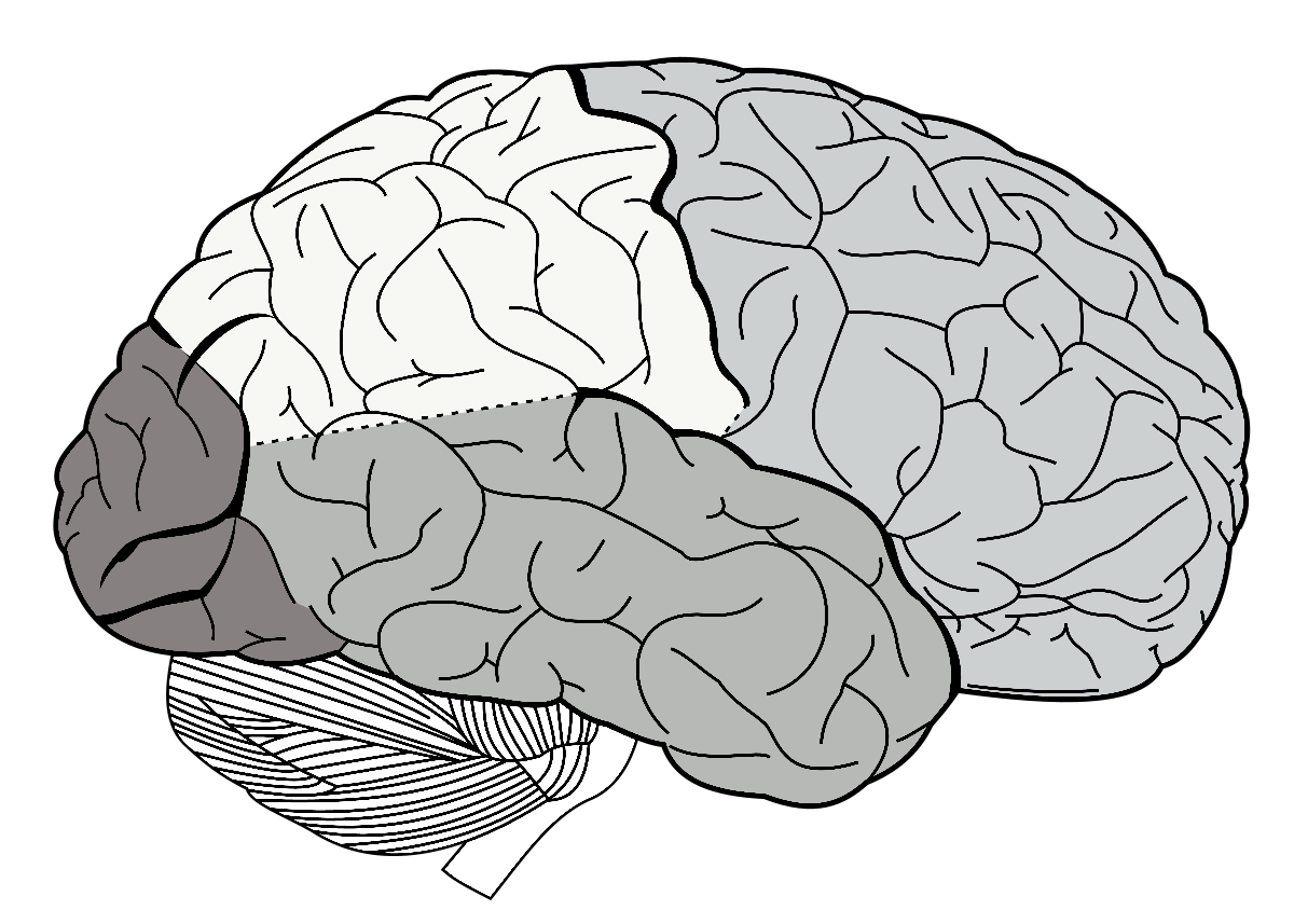
Cognitive neuroanatomy aims to explain how microanatomical structures generate macroanatomical functions contributing to measurable behaviors. At the heart of cognitive neuroanatomy is the goal to derive linking functions that can relate (a) behavioral output to (b) functional organization of the brain across spatial scales to (c) anatomical organization across spatial scales.
Electroencephalography (EEG) - Quantitative electroencephalography (QEEG) - Stereoelectroencephalography (SEEG) - Functional magnetic resonance imaging (fMRI) - Magnetoencephalography (MEG) - Near-infrared spectroscopy (NIRS) - Positron emission tomography (PET) - Single-unit recording (SUR) - Transcranial direct-current stimulation (TDCS) - Transcranial magnetic stimulation (TMS)
Electroencephalography (EEG) is an electrophysiological monitoring method to record electrical activity of the brain.
Theory: It is typically noninvasive, with the electrodes placed along the scalp, although invasive electrodes are sometimes used, as in electrocorticography. EEG measures voltage fluctuations resulting from ionic current within the neurons of the brain. Clinically, EEG refers to the recording of the brain's spontaneous electrical activity over a period of time, as recorded from multiple electrodes placed on the scalp.[1] Diagnostic applications generally focus either on event-related potentials or on the spectral content of EEG. The former investigates potential fluctuations time locked to an event, such as 'stimulus onset' or 'button press'. The latter analyses the type of neural oscillations (popularly called "brain waves") that can be observed in EEG signals in the frequency domain.
Applications: EEG is most often used to diagnose epilepsy, which causes abnormalities in EEG readings.[2] It is also used to diagnose sleep disorders, depth of anesthesia, coma, encephalopathies, and brain death. EEG used to be a first-line method of diagnosis for tumors, stroke and other focal brain disorders,[3][4] but this use has decreased with the advent of high-resolution anatomical imaging techniques such as magnetic resonance imaging (MRI) and computed tomography (CT). Despite limited spatial resolution, EEG continues to be a valuable tool for research and diagnosis. It is one of the few mobile techniques available and offers millisecond-range temporal resolution which is not possible with CT, PET or MRI. Derivatives of the EEG technique include evoked potentials (EP), which involves averaging the EEG activity time-locked to the presentation of a stimulus of some sort (visual, somatosensory, or auditory). Event-related potentials (ERPs) refer to averaged EEG responses that are time-locked to more complex processing of stimuli; this technique is used in cognitive science, cognitive psychology, and psychophysiological research.
Quantitative electroencephalography (QEEG) is a field concerned with the numerical analysis of electroencephalography (EEG) data and associated behavioral correlates.
Theory: Techniques used in digital signal analysis are extended to the analysis of electroencephalography (EEG); these include wavelet analysis and Fourier analysis, with new focus on shared activity between rhythms including phase synchrony (coherence, phase lag) and magnitude synchrony (comodulation/correlation, and asymmetry). The analog signal comprises a microvoltage time series of the EEG, sampled digitally and sampling rates adequate to over-sample the signal (using the Nyquist principle of exceeding twice the highest frequency being detected). Modern EEG amplifiers use adequate sampling to resolve the EEG across the traditional medical band from DC to 70 or 100 Hz, using sample rates of 250/256, 500/512, to over 1000 samples per second, depending on the intended application.
Applications: QEEG has been accepted for clinical application in some areas, such as cerebro-vascular disorders and epilepsy, though it remains yet to be accepted in other clinical areas, such as diagnosing mild traumatic brain injury or psychiatric disorders; the use of QEEG techniques in investigations in clinical and research settings are on going. QEEG has also been utilized to provide neurofeedback, which is a form of biofeedback, wherein electrical activity in the brain is monitored by a computer program, which is applied to modulate visual or auditory stimuli - These stimuli, in turn, are designed to be controlled by the user.
Stereoelectroencephalography (SEEG) is the practice of recording electroencephalographic signals via depth electrodes (electrodes surgically implanted into the brain tissue).
Theory: This technique was introduced in the diagnostic work up of patients with epilepsy by the group of the S. Anne Hospital, Paris, France, in the second half of the 20th century. Intracerebral electrodes are placed within the desired brain areas to record the electrical activity during epileptic seizures, thus contributing to define with accuracy the boundaries of the "epileptogenic zone", i.e. the area of brain generating the seizures which should be eventually surgically resected to achieve freedom from epileptic seizures. Potential risks of the procedure, accounting for less than 1% of cases, include brain hemorrhage and infection, which can lead to permanent neurological impairment or death. For this reason, stereoelectroencephalography is reserved to selected and particularly complicated epilepsy cases.
Applications: It may be used in patients with epilepsy not responding to medical treatment, and who are potential candidates to receive brain surgery in order to control seizures, it may also be used in research to collect neural data from specific regions of the brain, such as from the auditory cortex for auditory stimulus reconstruction.
Functional magnetic resonance imaging (fMRI) measures brain activity by detecting changes associated with blood flow.
Theory: This technique relies on the fact that cerebral blood flow and neuronal activation are coupled; when an area of the brain is in use, blood flow to that region also increases. The primary form of fMRI uses the blood-oxygen-level dependent (BOLD) contrast,[4] discovered by Seiji Ogawa; this is a type of specialized brain and body scan used to map neural activity in the brain or spinal cord of humans or other animals by imaging the change in blood flow (hemodynamic response) related to energy use by brain cells.
Applications:
Magnetoencephalography (MEG) is a functional neuroimaging technique for mapping brain activity by recording magnetic fields produced by electrical currents occurring naturally in the brain, using very sensitive magnetometers.
Theory: Arrays of SQUIDs (superconducting quantum interference devices) are currently the most common magnetometer, while the SERF (spin exchange relaxation-free) magnetometer is being investigated for future machines.
Applications: Applications of MEG include basic research into perceptual and cognitive brain processes, localizing regions affected by pathology before surgical removal, determining the function of various parts of the brain, and neurofeedback; this can be applied in a clinical setting to find locations of abnormalities as well as in an experimental setting to simply measure brain activity.
Near-infrared spectroscopy (NIRS) is a spectroscopic method that uses the near-infrared region of the electromagnetic spectrum (from 780 nm to 2500 nm).
Theory: Near-infrared spectroscopy is based on molecular overtone and combination vibrations; such transitions are forbidden by the selection rules of quantum mechanics. As a result, the molar absorptivity in the near-IR region is typically quite small.[citation needed] One advantage is that NIR can typically penetrate much further into a sample than mid infrared radiation. Near-infrared spectroscopy is, therefore, not a particularly sensitive technique, but it can be very useful in probing bulk material with little or no sample preparation. The molecular overtone and combination bands seen in the near-IR are typically very broad, leading to complex spectra; it can be difficult to assign specific features to specific chemical components. Multivariate (multiple variables) calibration techniques (e.g., principal components analysis, partial least squares, or artificial neural networks) are often employed to extract the desired chemical information. Careful development of a set of calibration samples and application of multivariate calibration techniques is essential for near-infrared analytical methods.
Applications; Typical applications include medical and physiological diagnostics and research including blood sugar, pulse oximetry, functional neuroimaging, sports medicine, elite sports training, ergonomics, rehabilitation, neonatal research, brain computer interface, urology (bladder contraction), and neurology (neurovascular coupling). There are also applications in other areas as well such as pharmaceutical, food and agrochemical quality control, atmospheric chemistry, combustion research and astronomy.
Positron-emission tomography (PET) is a nuclear medicine functional imaging technique that is used to observe metabolic processes in the body as an aid to the diagnosis of disease.
Theory: The system detects pairs of gamma rays emitted indirectly by a positron-emitting radioligand, most commonly fluorine-18, which is introduced into the body on a biologically active molecule called a radioactive tracer.
Applications: Different ligands are used for different imaging purposes, depending on what the radiologist/researcher wants to detect. Three-dimensional images of tracer concentration within the body are then constructed by computer analysis. In modern PET computed tomography scanners, three-dimensional imaging is often accomplished with the aid of a computed tomography X-ray scan performed on the patient during the same session, in the same machine. If the biologically active tracer molecule chosen for PET is fludeoxyglucose (FDG), an analogue of glucose, the concentrations of tracer imaged will indicate tissue metabolic activity as it corresponds to the regional glucose uptake. Use of this tracer to explore the possibility of cancer metastasis (i.e., spreading to other sites) is the most common type of PET scan in standard medical care (representing 90% of current scans). Metabolic trapping of the radioactive glucose molecule allows the PET scan to be utilized;[2] the same tracer may also be used for PET investigation and diagnosis of types of dementia. Less often, other radioactive tracers, usually but not always labeled with fluorine-18, are used to image the tissue concentration of other types of molecules of interest. One of the disadvantages of PET scanners is their operating cost.[3] A similar imaging process to PET is single-photon emission computed tomography (SPECT), which also uses radioligands to detect molecules in the brain, and is less expensive.
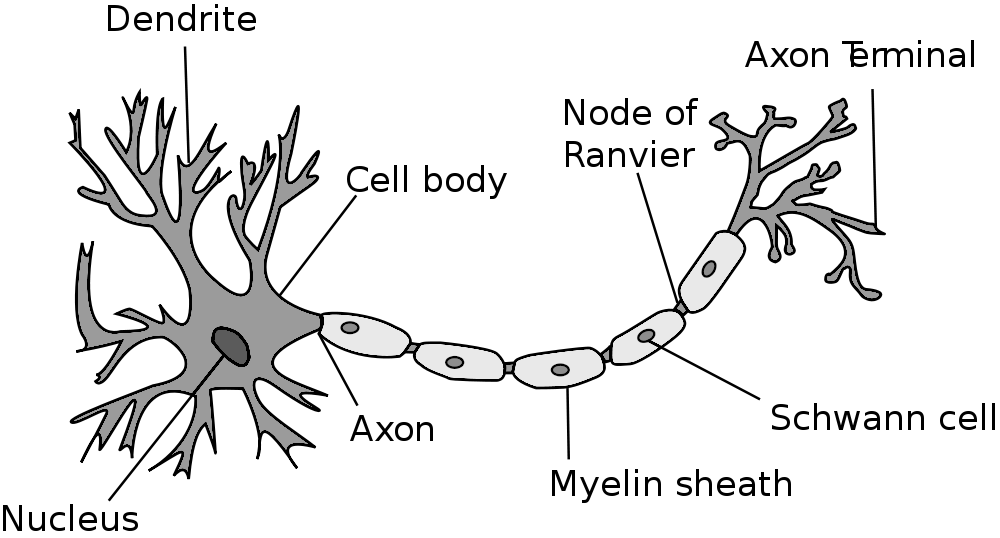
Single-unit recordings provide a method of measuring the electro-physiological responses of single neurons using a microelectrode system.
Theory: When a neuron generates an action potential, the signal propagates down the neuron as a current which flows in and out of the cell through excitable membrane regions in the soma and axon. A microelectrode is inserted into the brain, where it can record the rate of change in voltage with respect to time. These microelectrodes must be fine-tipped, high-impedance conductors; they are primarily glass micro-pipettes or metal microelectrodes made of platinum or tungsten.[1] Microelectrodes can be carefully placed close to the cell membrane, allowing the ability to record extracellularly.
Applications: Single-unit recordings are widely used in cognitive science, where it permits the analysis of human cognition and cortical mapping. This information can then be applied to brain machine interface (BMI) technologies for brain control of external devices.
Transcranial direct current stimulation (tDCS) is a form of neuromodulation that uses constant, low direct current delivered via electrodes on the head. It can be contrasted with cranial electrotherapy stimulation, which generally uses alternating current the same way.
Theory: In transcranial magnetic stimulation (TMS), an electric coil is held above the region of interest on the scalp that uses rapidly changing magnetic fields to induce small electrical currents in the brain. There are two types of TMS: repetitive TMS and single pulse TMS. Both are used in research therapy but effects lasting longer than the stimulation period are only observed in repetitive TMS. Similar to tDCS, an increase or decrease in neuronal activity can be achieved using this technique, but the method of how this is induced is very different. Transcranial direct current stimulation has the two different directions of current that cause the different effects. Increased neuronal activity is induced in repetitive TMS by using a higher frequency and decreased neuronal activity is induced by using a lower frequency.
Applications: TDCS appears to be somewhat effective for depression.[4] There is also evidence that tDCS is useful in treating neuropathic pain after spinal cord injury [17] and improving activities of daily living assessment after stroke.
Transcranial magnetic stimulation (TMS), also known as repetitive transcranial magnetic stimulation (rTMS), is a noninvasive form of brain stimulation in which a changing magnetic field is used to cause electric current at a specific area of the brain through electromagnetic induction.
Theory: An electric pulse generator, or stimulator, is connected to a magnetic coil, which in turn is connected to the scalp. The stimulator generates a changing electric current within the coil which induces a magnetic field; this field then causes a second inductance of inverted electric charge within the brain itself.
Applications: TMS has shown diagnostic and therapeutic potential in the central nervous system with a wide variety of disease states in neurology and mental health, with research still evolving. Adverse effects of TMS are rare, and include fainting and seizure.[7] Other potential issues include discomfort, pain, hypomania, cognitive change, hearing loss, and inadvertent current induction in implanted devices such as pacemakers or defibrillators.
Computer Animations
Brain structure - Brodmann Areas (BA) - Skull bones - Cranial bones - Facial bones
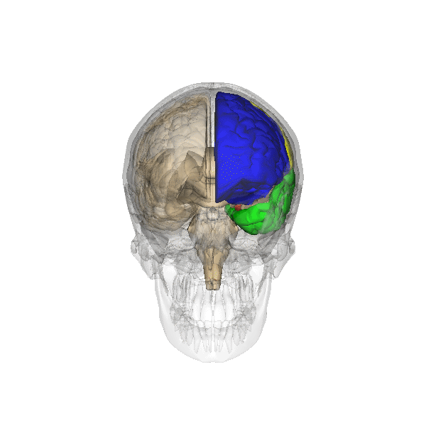
Figure 1. Four lobes (computer animation n°0). Link |
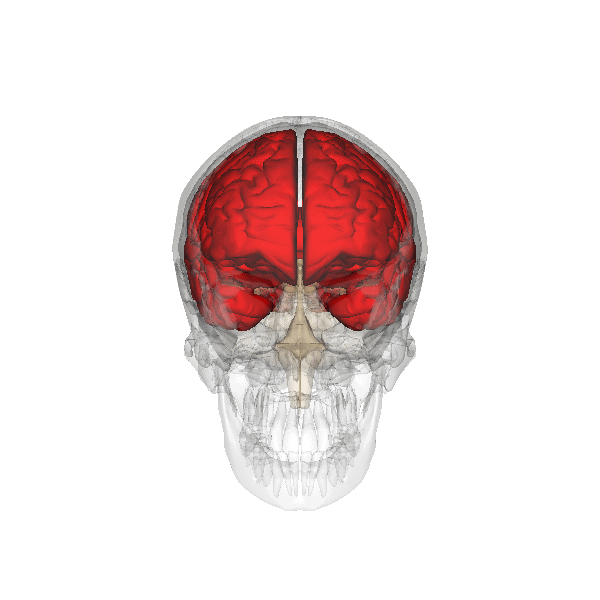
Figure 2. Telencephalon (computer animation n°0). Link |
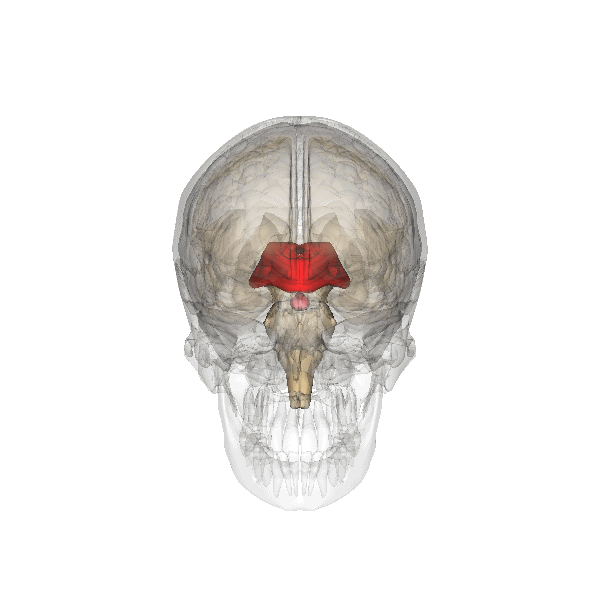
Figure 3. Diencephalon (computer animation n°0). Link |
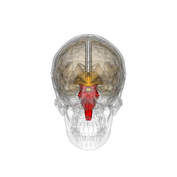
Figure 4. Brain stem (computer animation n°0). Link |

Figure 5. Cerebellum (computer animation n°0). Link |
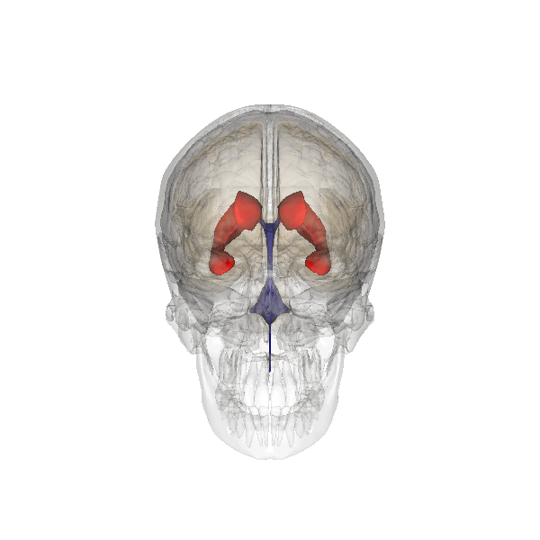
Figure 6. Lateral ventricle (computer animation n°0). Link |
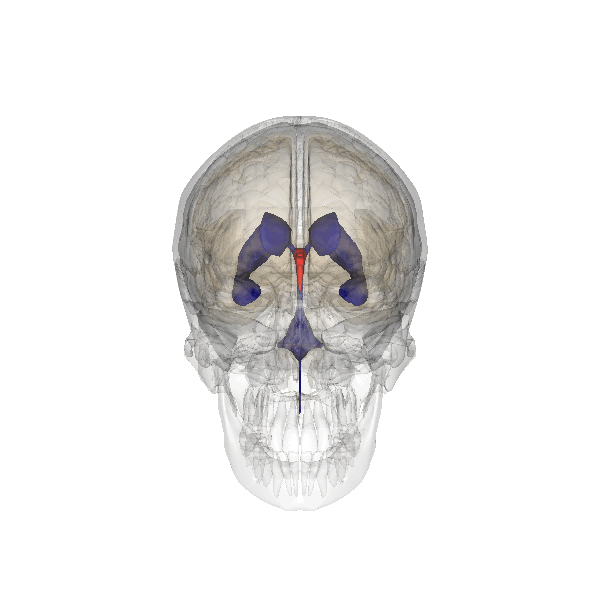
Figure 7. Third ventricle (computer animation n°0). Link |
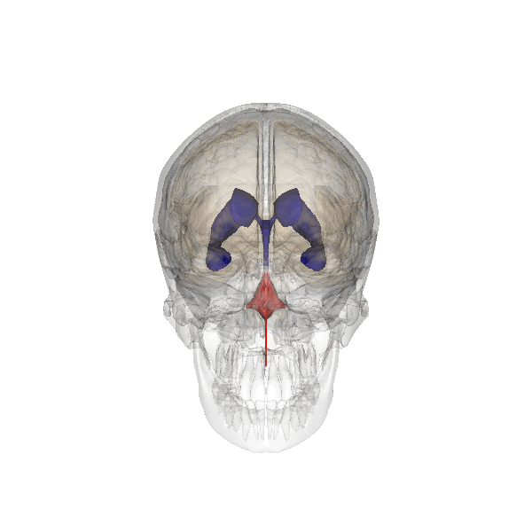
Figure 8. Fourth (computer animation n°0). Link |
|
Figure 9. Four lobes (computer animation n°0). Link |
Figure 10. Rotating brain (computer animation n°0). Link |
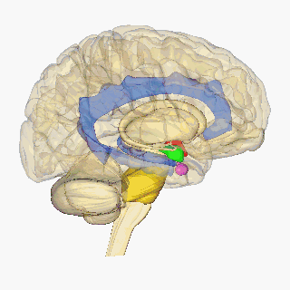
Figure 11. Rotating brain colored (computer animation n°0). Link |
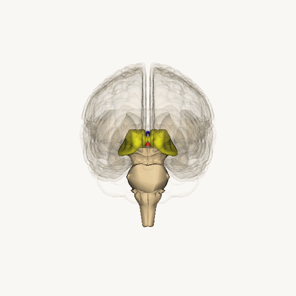
Figure 12. Rotating brain stem and thalamus (computer animation n°0). Link |
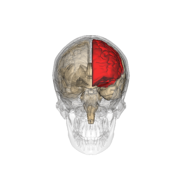
Figure 13. Frontal lobe (computer animation n°0). Link |

Figure 14. Occipital lobe (computer animation n°0). Link |
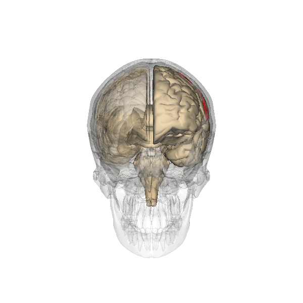
Figure 15. Parietal lobe (computer animation n°0). Link |
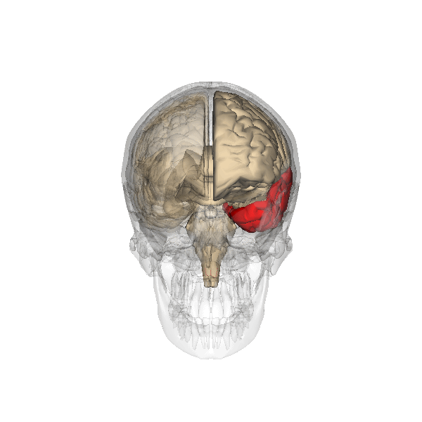
Figure 16. Temporal lobe (computer animation n°0). Link |
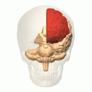
Figure 17. Frontal lobe (computer animation n°1). Link |

Figure 18. Occipital lobe (computer animation n°1). Link |
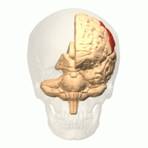
Figure 19. Parietal lobe (computer animation n°1). Link |
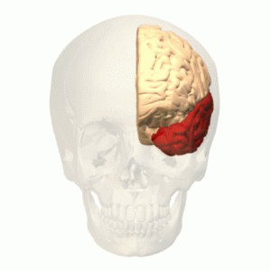
Figure 20. Temporal lobe (computer animation n°1). Link |
|
Figure 21. Brain stem (computer animation n°0). Link |
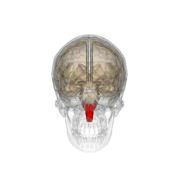
Figure 22. Medulla oblongata (computer animation n°0). Link |
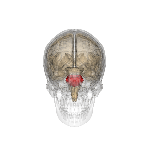
Figure 23. Pons (computer animation n°0). Link |
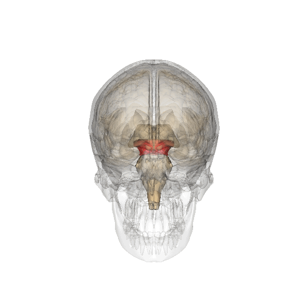
Figure 24. Midbrain (computer animation n°0). Link |

Figure 25. Limbic lobe (computer animation n°0). Link |
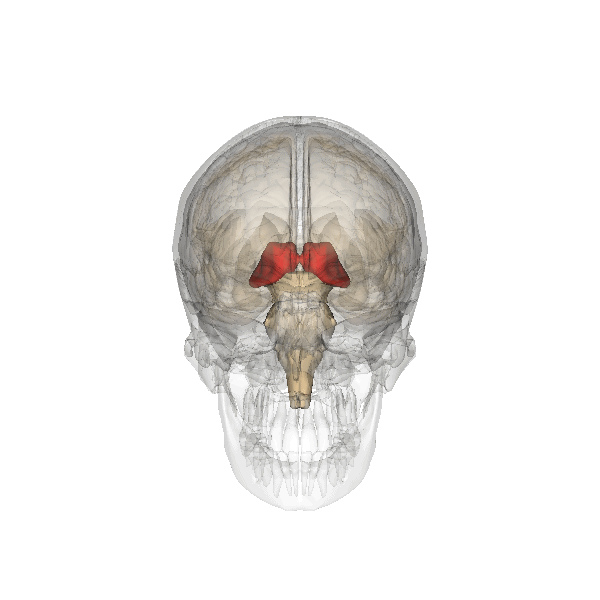
Figure 26. Thalamus (computer animation n°0). Link |
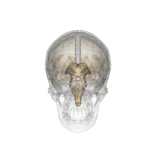
Figure 27. Pineal_gland (computer animation n°0). Link |
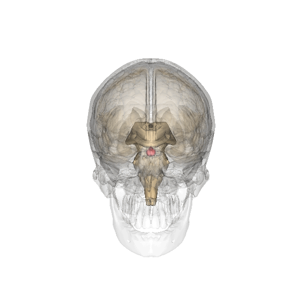
Figure 28. Pituitary_gland (computer animation n°0). Link |
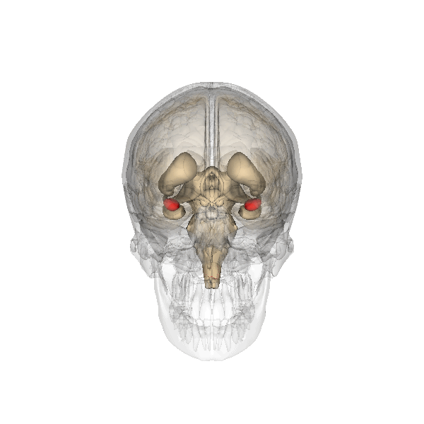
Figure 29. Amygdala (computer animation n°0). Link |

Figure 30. Corpus callosum (computer animation n°0). Link |
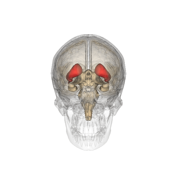
Figure 31. Striatum (caudate nucleus, putamen, internal capsule) (computer animation n°0). Link |
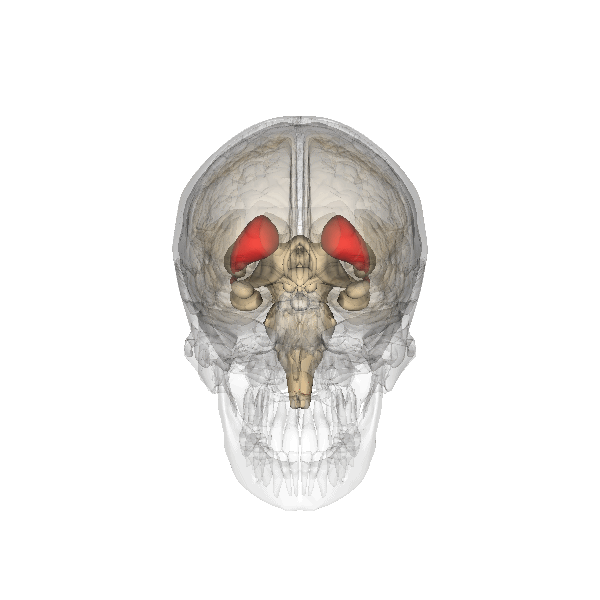
Figure 32. Caudate nucleus (computer animation n°0). Link |
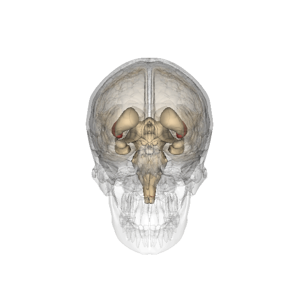
Figure 33. Putamen (computer animation n°0). Link |
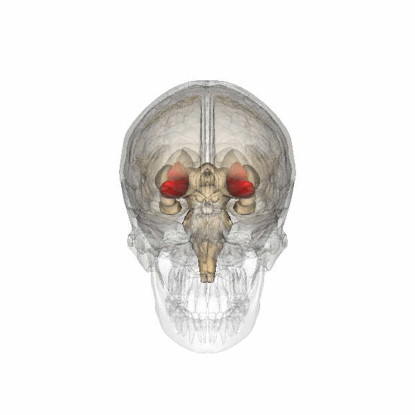
Figure 34. Internal capsule (computer animation n°0). Link |
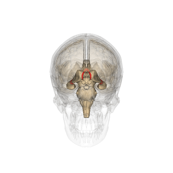
Figure 35. Fornix (computer animation n°0). Link |
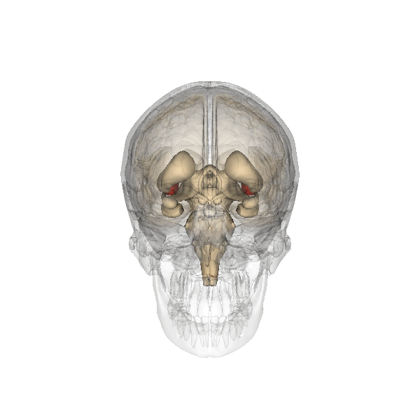
Figure 36. Globus pallidus (computer animation n°0). Link |

Figure 37. Inferior colliculus (computer animation n°0). Link |

Figure 38. Superior colliculus (computer animation n°0). Link |

Figure 39. Uncus (computer animation n°0). Link |
Figure 40. Dorsal and ventral pathways (computer animation n°0). Link |
.gif)
Figure 41. Phineas Gage injury (computer animation n°0). Link |
Figure 42. HPA axis (computer animation n°0). Link |
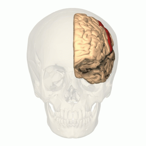
Figure 1. BA 3, 1, 2 (computer animation n°0). Link |
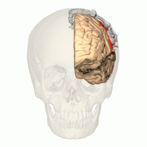
Figure 2. BA 3, 1, 2 with homunculus (computer animation n°0). Link |
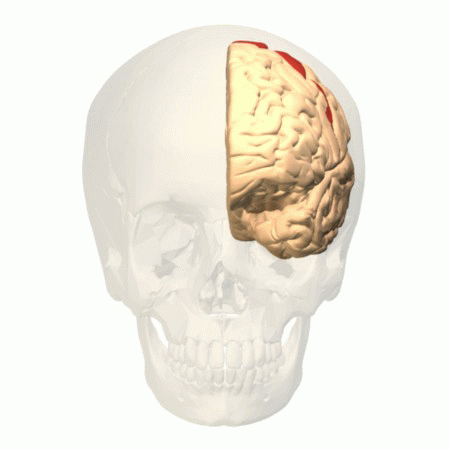
Figure 3. BA 4 (computer animation n°0). Link |
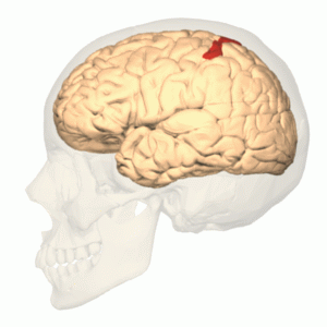
Figure 4. BA 5 (computer animation n°0). Link |
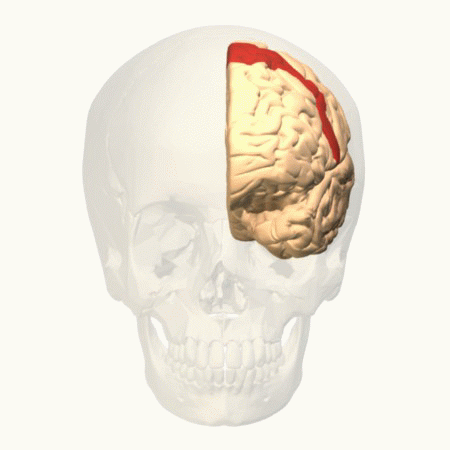
Figure 5. BA 6 (computer animation n°0). Link |
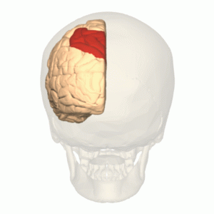
Figure 6. BA 7 (computer animation n°0). Link |
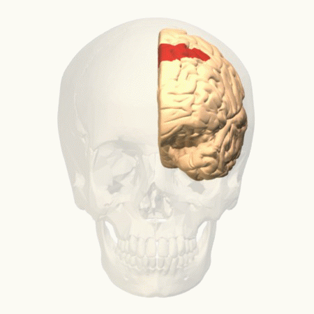
Figure 7. BA 8 (computer animation n°0). Link |
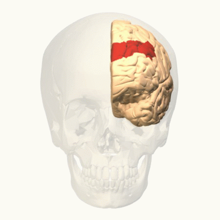
Figure 8. BA 9 (computer animation n°0). Link |
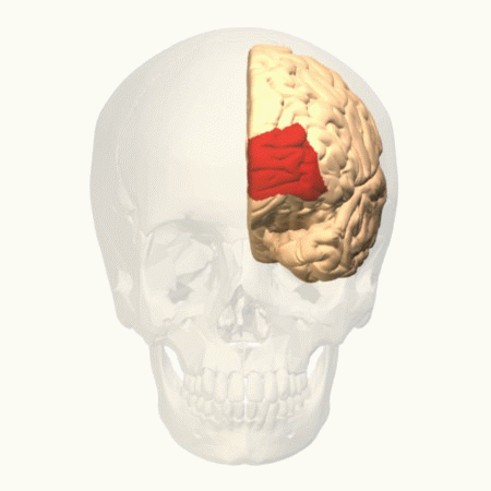
Figure 9. BA 10 (computer animation n°0). Link |

Figure 10. BA 11 (computer animation n°0). Link |

Figure 11. BA 12 (computer animation n°0). Link |
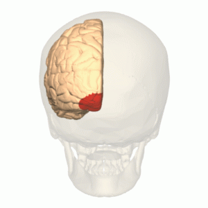
Figure 12. BA 17 (computer animation n°0). Link |
_-_animation.gif)
Figure 13. BA 18 (computer animation n°0). Link |
_-_animation.gif)
Figure 14. BA 19 (computer animation n°0). Link |
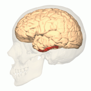
Figure 15. BA 20 (computer animation n°0). Link |
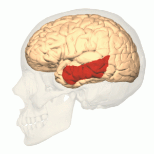
Figure 16. BA 21 (computer animation n°0). Link |

Figure 17. BA 22 (computer animation n°0). Link |

Figure 18. BA 24 (computer animation n°0). Link |

Figure 19. BA 25 (computer animation n°0). Link |
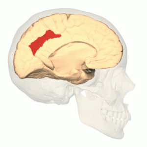
Figure 20. BA 31 (computer animation n°0). Link |

Figure 21. BA 32 (computer animation n°0). Link |
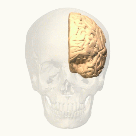
Figure 22. BA 33 (computer animation n°0). Link |
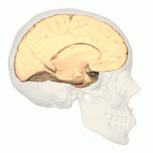
Figure 23. BA 35 (computer animation n°0). Link |
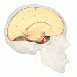
Figure 24. BA 36 (computer animation n°0). Link |
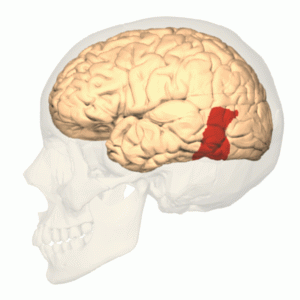
Figure 25. BA 37 (computer animation n°0). Link |
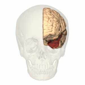
Figure 26. BA 38 (computer animation n°0). Link |
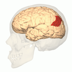
Figure 27. BA 39 (computer animation n°0). Link |
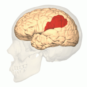
Figure 28. BA 40 (computer animation n°0). Link |
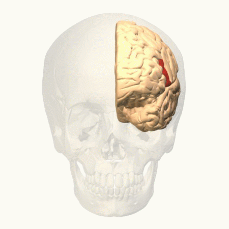
Figure 29. BA 44 (computer animation n°0). Link |
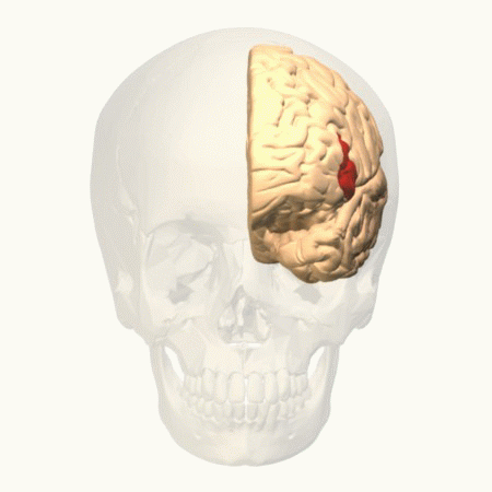
Figure 30. BA 45 (computer animation n°0). Link |
Figure 310. BA 46 (computer animation n°0). Link |
Figure 32. BA 47 (computer animation n°0). Link |
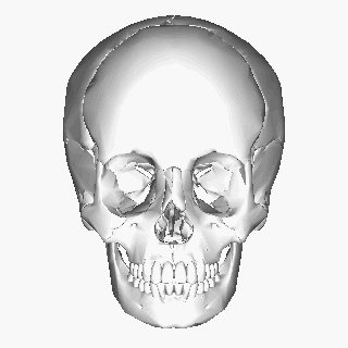
Figure 1. Human skull (computer animation n°1). Link |
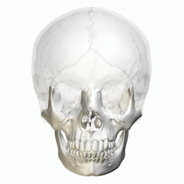
Figure 2. Neurocranium (computer animation N°0). Link |
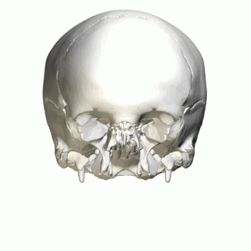
Figure 3. Neurocranium (computer animation N°4). Link |
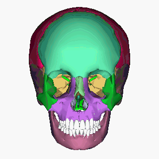
Figure 4. Human skull (computer animation N°2). Link |
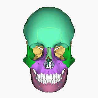
Figure 5. Human skull (computer animation N°3). Link |
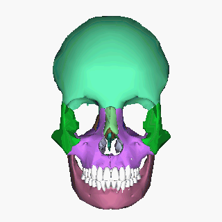
Figure 6. Human skull (computer animation N°4). Link |
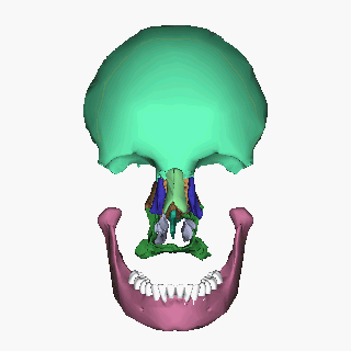
Figure 8. Human skull (computer animation N°5). Link |
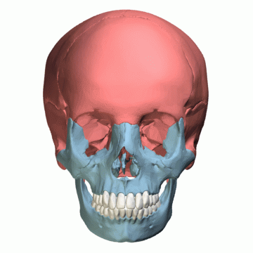
Figure 9. Facial bones and Cranial bones (computer animation N°1). Link |
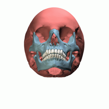
Figure 10. Facial bones and Cranial bones (computer animation N°2). Link |
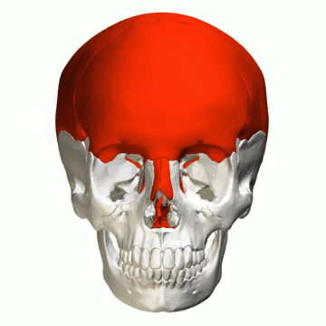
Figure 11. Flat_bones in skull (computer animation). Link |
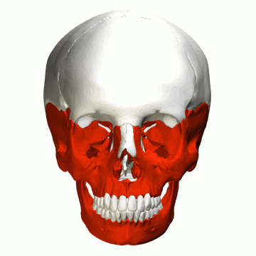
Figure 12. Irregular bones in_skull (computer animation). Link |
|
Figure 1. Human skull (computer animation N°1). Link |
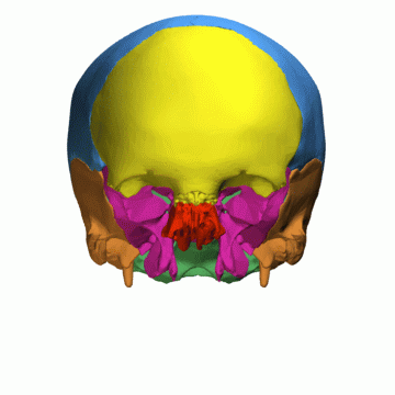
Figure 2. Neurocranium (computer animation N°1). Link |
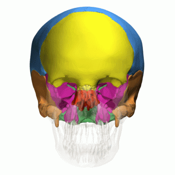
Figure 3. Neurocranium (computer animation N°2). Link |
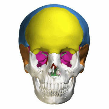
Figure 4. Neurocranium (computer animation N°3). Link |
|
Figure 5. Neurocranium (computer animation N°4). Link |
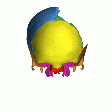
Figure 6. Neurocranium, superior view (computer animation n°1). Link |
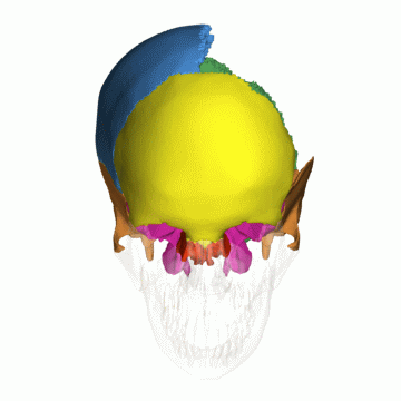
Figure 7. Neurocranium, superior view (computer animation n°2). Link |
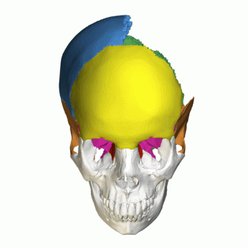
Figure 8. Neurocranium : superior view (computer animation n°3). Link |
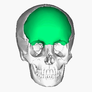
Figure 9. Frontal bone (computer animation N°0). Link |
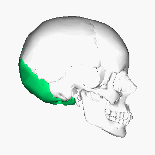
Figure 10. Occipital bone (computer animation N°0). Link |
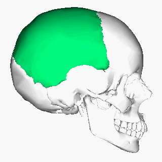
Figure 11. Parietal bone (computer animation N°0). Link |
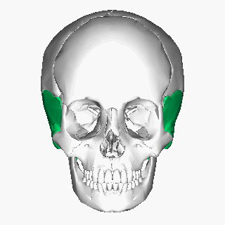
Figure 12. Temporal bone : lateral view (computer animation N°0). Link |
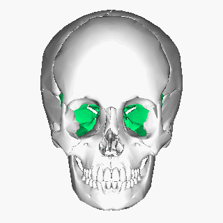
Figure 13. Sphenoid bone (computer animation N°1). Link |

Figure 14. Sphenoid bone : inferior view (computer animation N°2). Link |
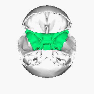
Figure 15.Sphenoid bone : superior view (computer animation N°3). Link |
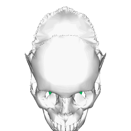
Figure 16. Sphenoid bone : superior view (computer animation N°4). Link |
|
Figure 17. Neurocranium (computer animation N°2). Link |
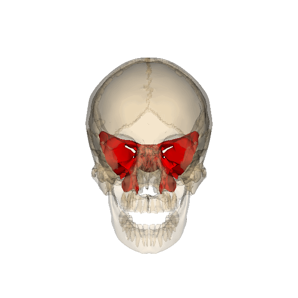
Figure 18. Sphenoid bone : rotation (computer animation n°0). Link |
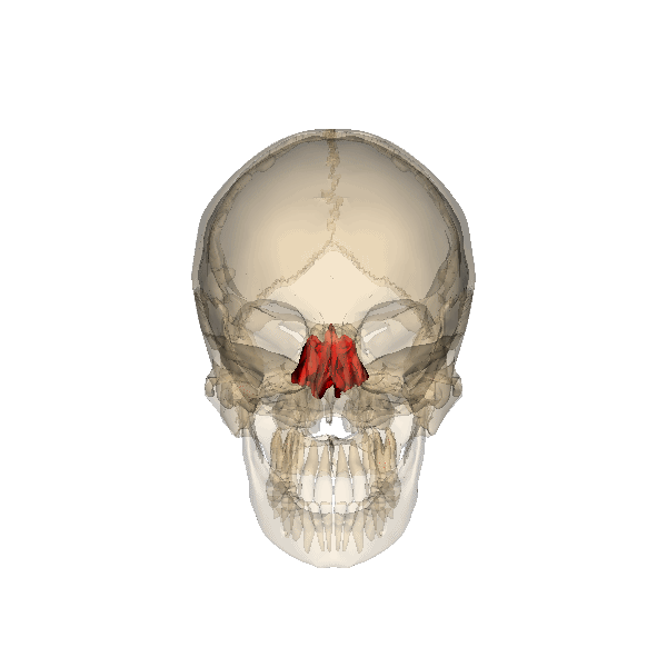
Figure 19. Ethmoid bone : rotation (computer animation N°0). Link |
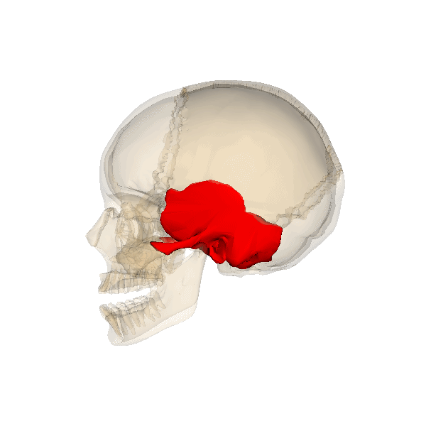
Figure 20. Temporal bone rotation (computer animation N°1). Link |
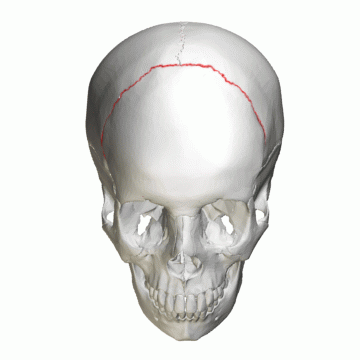
Figure 21. Coronal suture (computer animation N°1). Link |
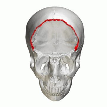
Figure 22. Coronal suture (computer animation N°2). Link |
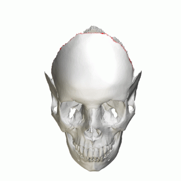
Figure 23. Coronal suture (computer animation N°3). Link |
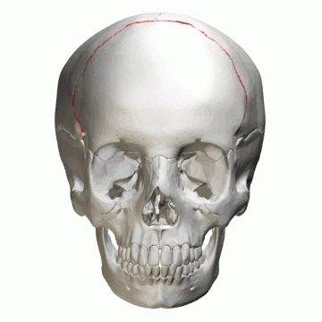
Figure 24. Coronal suture (computer animation N°4). Link |
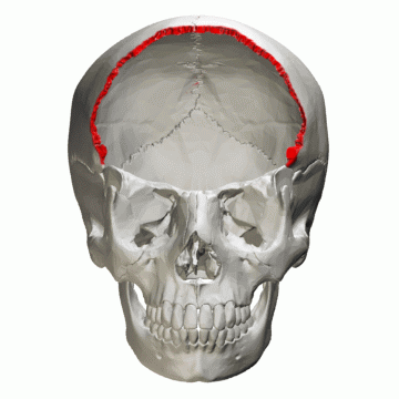
Figure 25. Coronal suture (computer animation N°5). Link |
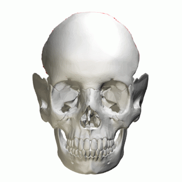
Figure 26. Coronal suture (computer animation N°6). Link |
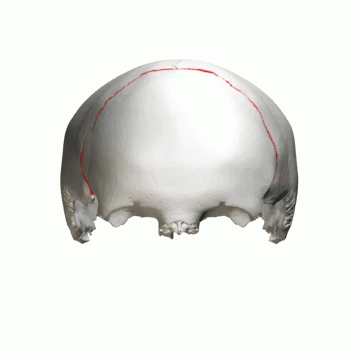
Figure 27. Coronal suture (computer animation N°7). Link |
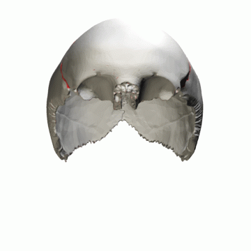
Figure 28. Coronal suture (computer animation N°8). Link |
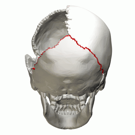
Figure 29. Lambdoid suture (computer animation N°5). Link |
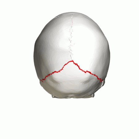
Figure 30. Lambdoid suture (computer animation N°6). Link |
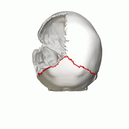
Figure 31. Lambdoid suture (computer animation N°7). Link |
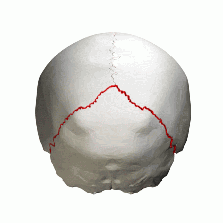
Figure 32. Lambdoid suture (computer animation N°8). Link |
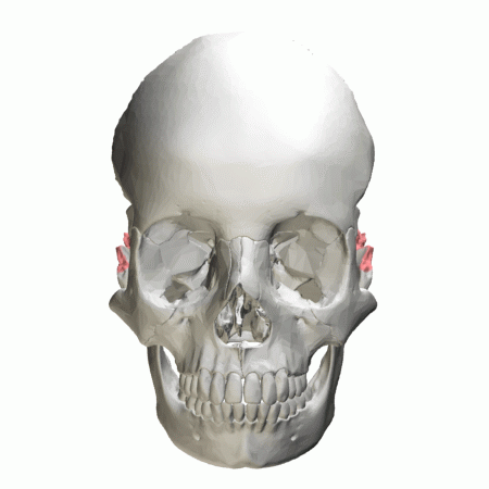
Figure 33. Lambdoid border of occipital bone (computer animation N°5). Link |
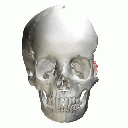
Figure 34. Lambdoid border of occipital bone (computer animation N°6). Link |
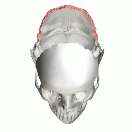
Figure 35. Lambdoid border of occipital bone (computer animation N°7). Link |
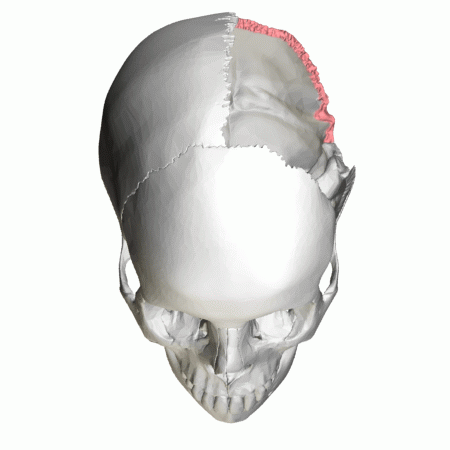
Figure 36. Lambdoid border of occipital bone (computer animation N°8). Link |

Figure 37. Sphenoparietal suture (computer animation N°1). Link |
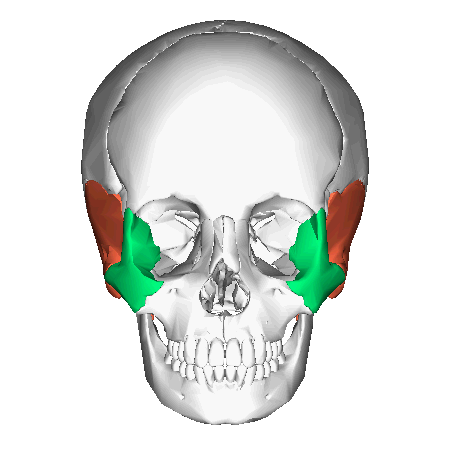
Figure 38. Zygomaticotemporal suture (computer animation N°2). Link |
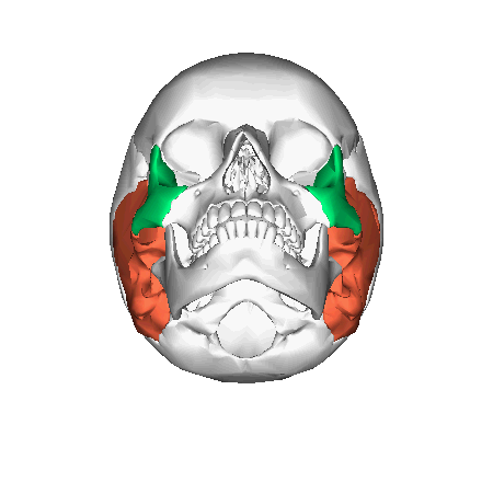
Figure 39. Zygomaticotemporal suture : inferior view (computer animation N°3). Link |
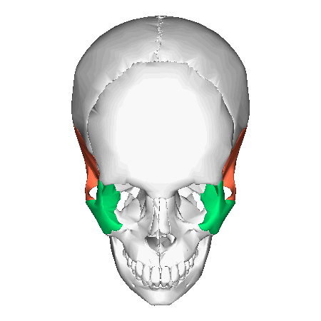
Figure 40. Zygomaticotemporal suture : superior view (computer animation N°4). Link |
|
Figure 41. Frontal bone (computer animation N°0). Link |
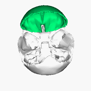
Figure 42. Frontal bone (computer animation N°2). Link |
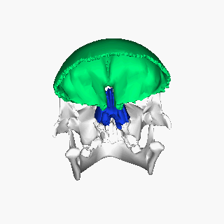
Figure 43. Frontal bone (computer animation N°3). Link |
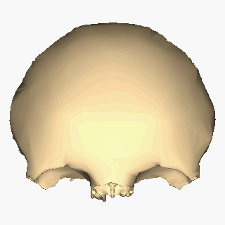
Figure 44. Frontal bone close-up (computer animation N°4). Link |
|
Figure 45. Occipital bone (computer animation N°0). Link |
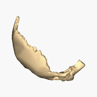
Figure 46. Occipital bone close-up : lateral view (computer animation N°1). Link |
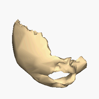
Figure 47. Occipital bone close-up : inferior view (computer animation N°2). Link |
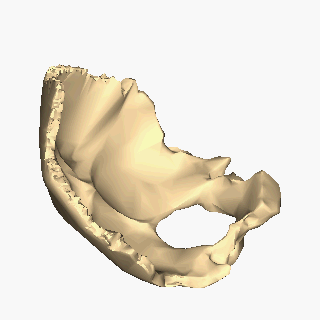
Figure 48. Occipital bone close-up : superior view (computer animation N°3). Link |
|
Figure 49. Parietal bone (computer animation N°0). Link |
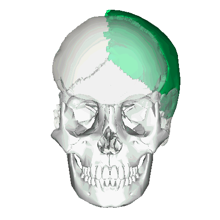
Figure 50. Left parietal bone (computer animation N°0). Link |
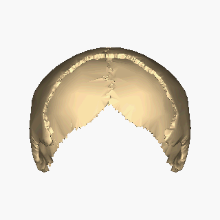
Figure 51. Parietal bone close-up (computer animation N°1). Link |
Figure 52. Parietal bone close-up (computer animation N°2). Link |
|
Figure 53. Temporal bone : lateral view (computer animation N°0). Link |
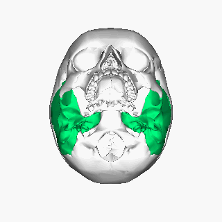
Figure 54. Temporal bone : inferior view (computer animation N°1). Link |
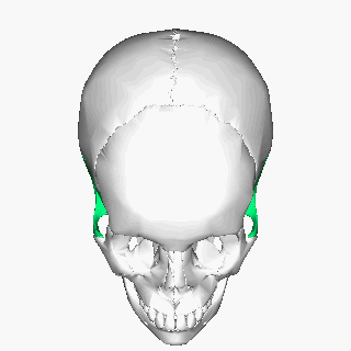
Figure 55. Temporal bone : superior view (computer animation N°2). Link |
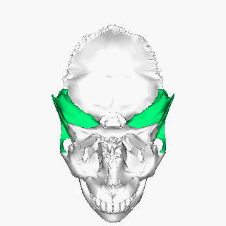
Figure 56. Temporal bone : superior view (computer animation N°3). Link |
|
Figure 57. Sphenoid bone (computer animation N°1). Link |
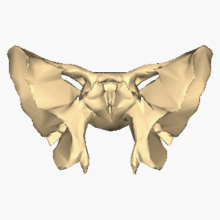
Figure 58. Sphenoid bone close-up (computer animation N°1). Link |
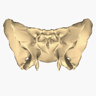
Figure 59. Sphenoid bone close-up : inferior view (computer animation N°2). Link |
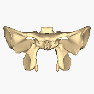
Figure 60. Sphenoid bone close-up : superior view (computer animation N°3). Link |
|
Figure 61. Sphenoid bone (computer animation N°1). Link |
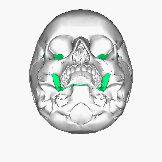
Figure 62. Sphenoid bone : inferior view (computer animation N°2). Link |
Figure 63. Sphenoid bone : superior view (computer animation N°3). Link |
Figure 64. Sphenoid bone : superior view (computer animation N°4). Link |
|
Figure 1. Neurocranium (computer animation N°0). Link |
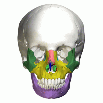
Figure 2. Facial bones (computer animation n°1). Link |
Figure 3. Facial bones and Cranial bones (computer animation N°1). Link |
Figure 4. Facial bones and Cranial bones (computer animation N°2). Link |
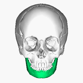
Figure 5. Mandible (computer animation N°0). Link |
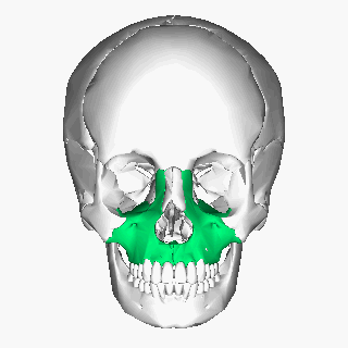
Figure 6. Maxilla (computer animation n°0). Link |
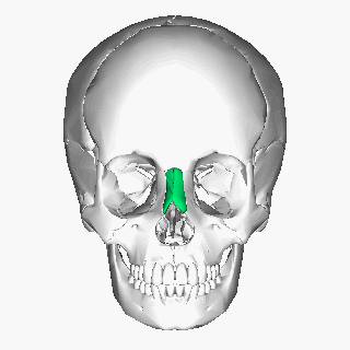
Figure 7. Nasal bone (computer animation n°0). Link |
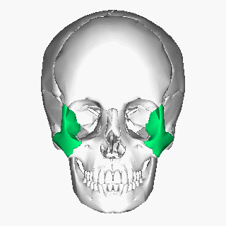
Figure 8. Zygomatic bone : lateral view (computer animation n°0). Link |
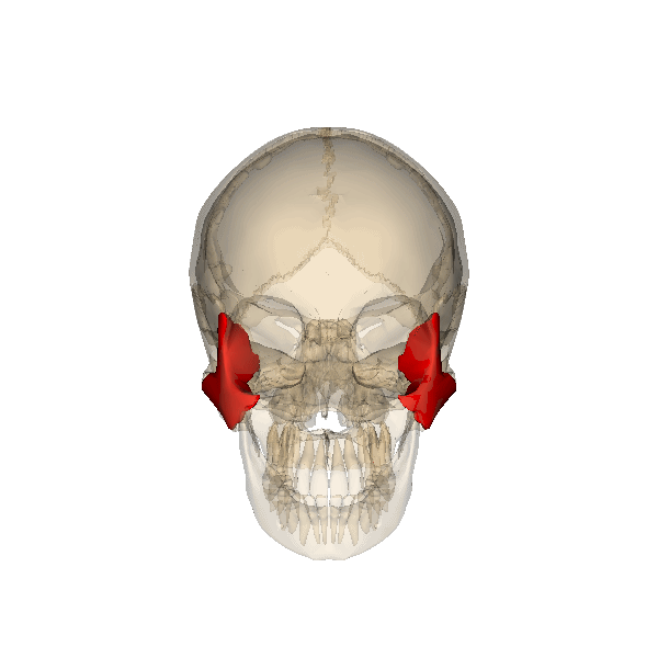
Figure 9. Zygomatic bone : rotation (computer animation N°0). Link |
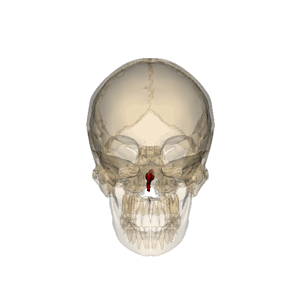
Figure 10. Vomer bone : rotation (computer animation N°0). Link |
Figure 11. Lacrimal bone (computer animation N°1). Link |
Figure 12. Lacrimal bone : inferior view (computer animation N°2). Link |
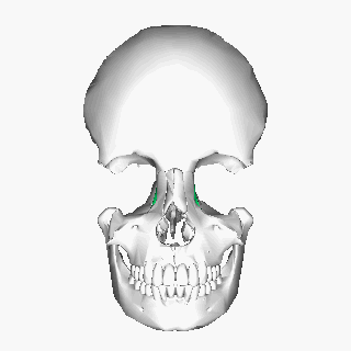
Figure 13. Lacrimal bone : superior view (computer animation N°3). Link |
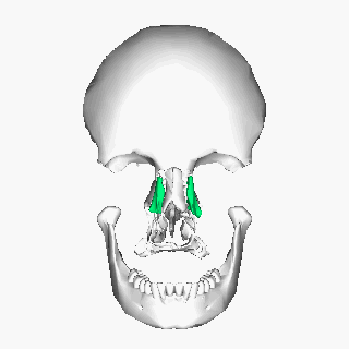
Figure 14. Lacrimal : superior view (computer animation N°4). Link |
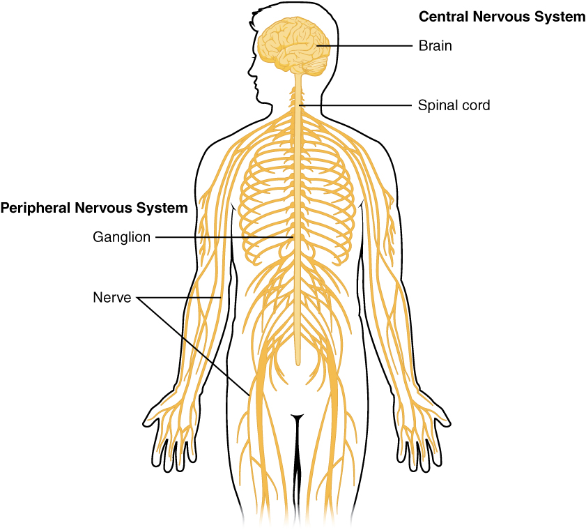
Figure 1. Schematic diagram showing the central nervous system in yellow, peripheral in orange (from Wikipedia: OpenStax Anatomy and Physiology). Link |
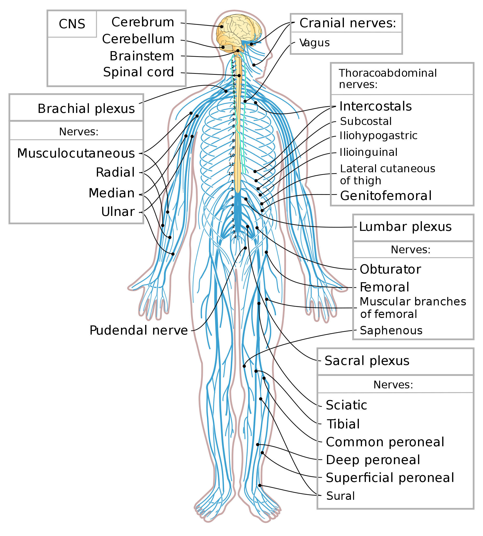
Figure 2. Schematic diagram showing the central nervous system in yellow, peripheral in blue (from Wikipedia). Link |
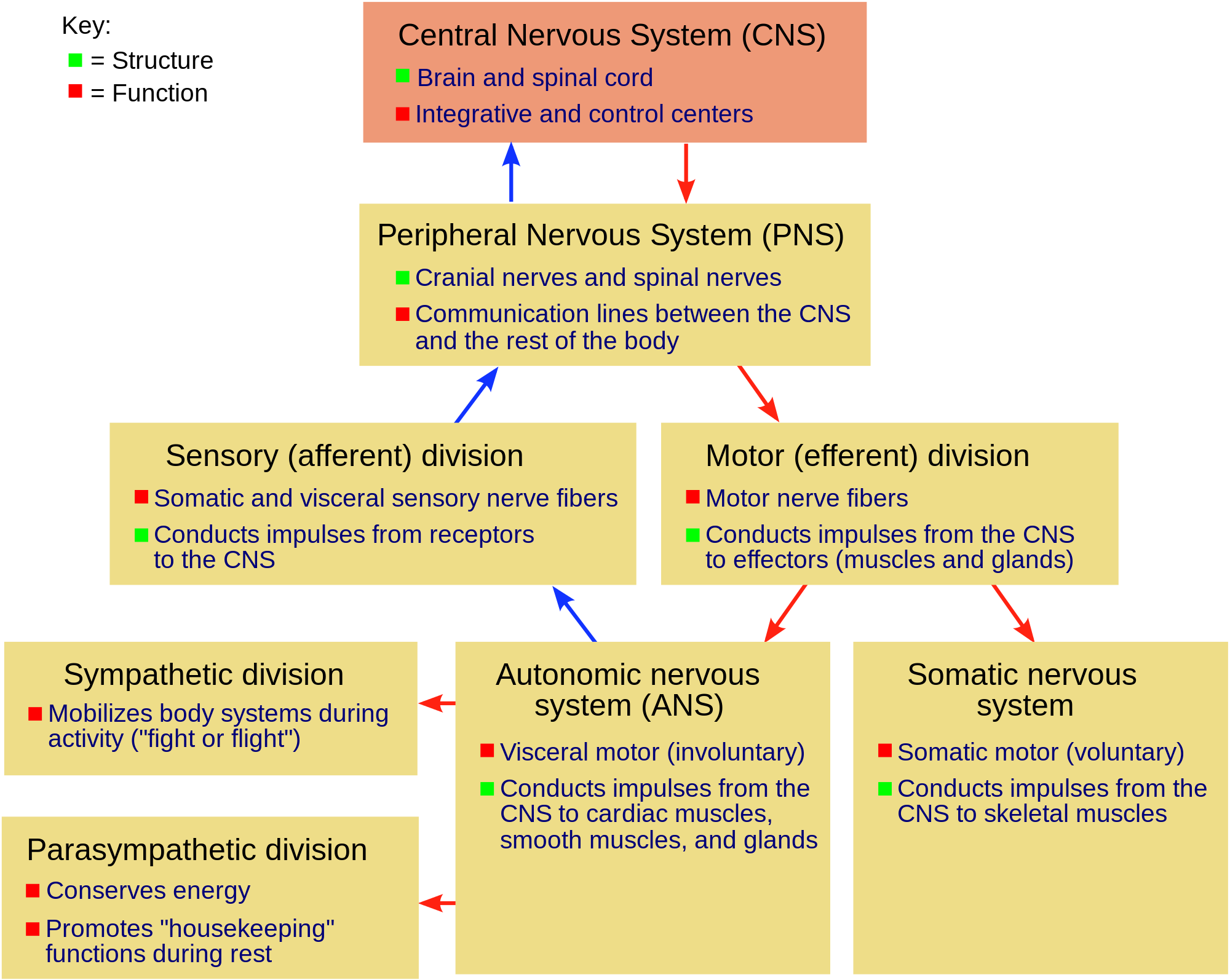
Figure 3. Diagram showing the major divisions of the vertebrate nervous system (from Wikipedia). Link |
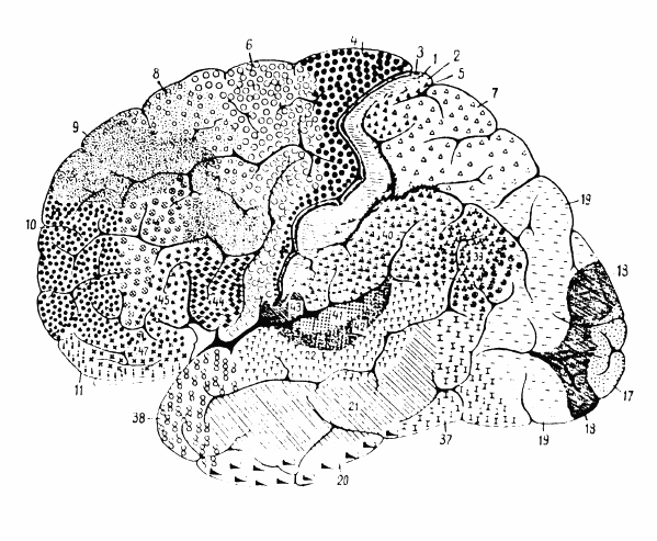
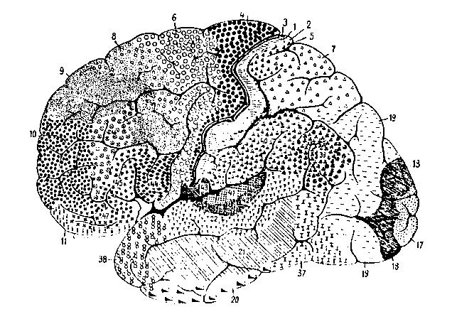
Figure 1. Cytoarchitectonics of human brain according to Brodmann (1909) : lateral surface (from Brodmann, K. (1909). Vergleichende Lokalisationslehre der Grosshirnrinde in ihren Prinzipien dargestellt auf Grund des Zellenbaues. Johann Ambrosius Barth Verlag, Leipz). Link |
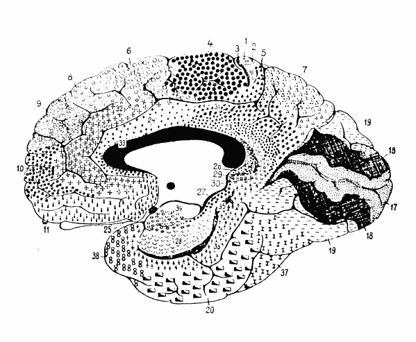
Figure 2. Cytoarchitectonics of human brain according to Brodmann (1909) : medial surface (from Brodmann, K. (1909). Vergleichende Lokalisationslehre der Grosshirnrinde in ihren Prinzipien dargestellt auf Grund des Zellenbaues. Johann Ambrosius Barth Verlag, Leipz). Link |
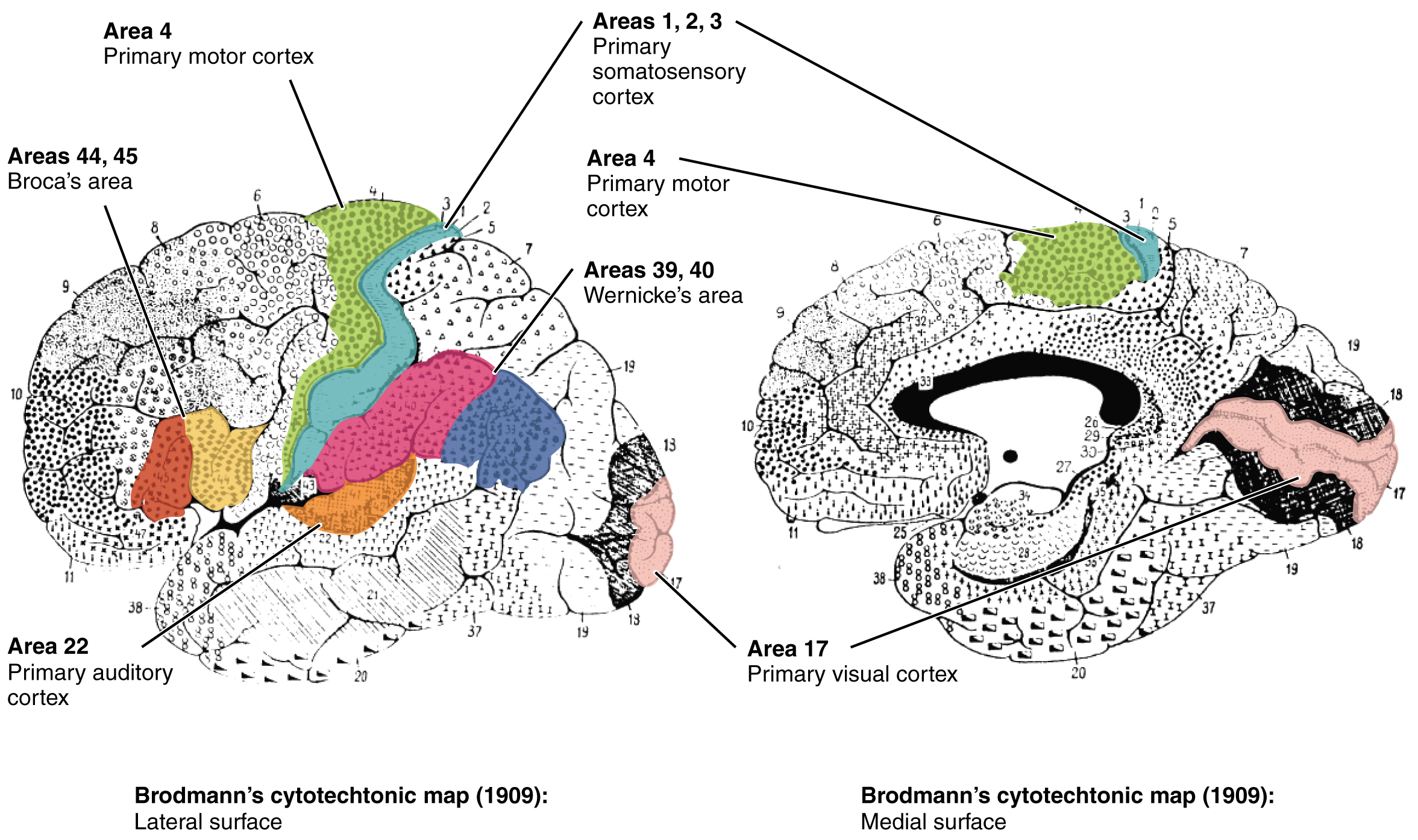
Figure 3. Cytoarchitectonics of human brain to Brodmann (1909) (colored) : lateral surface and medial surface (from Wikipedia: OpenStax Anatomy and Physiology). Link |
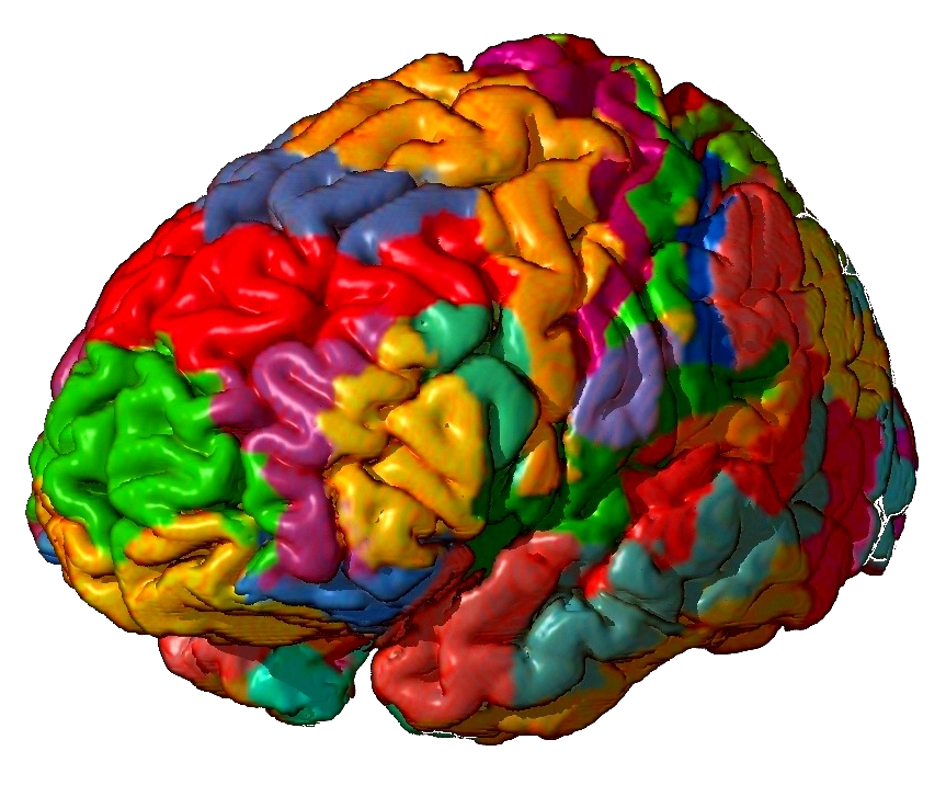
Figure 4. 3D representation of Brodmann areas (view 1) (from (from Wikipedia: Mark Dow.) Cytoarchitectonics of human brain (colored) : lateral surface (from Wikipedia: OpenStax Anatomy and Physiology). Link |
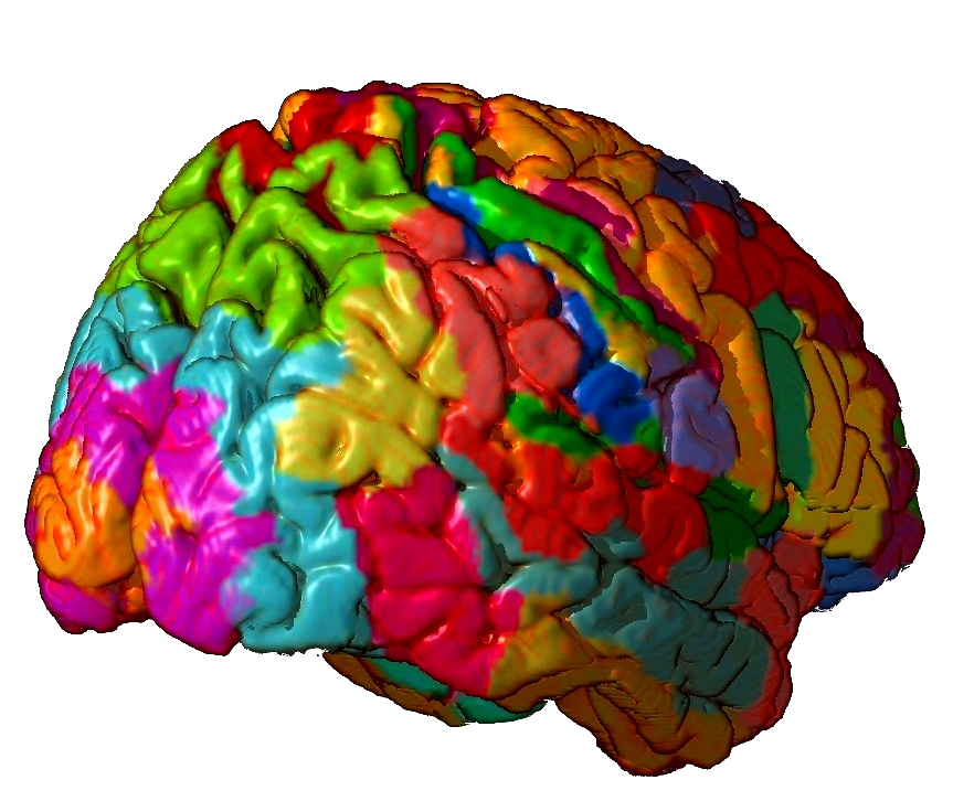
Figure 5. 3D representation of Brodmann areas (view 2) (from (from Wikipedia: Mark Dow.)Cytoarchitectonics of human brain (colored) : medial surface (from Wikipedia: OpenStax Anatomy and Physiology). Link |
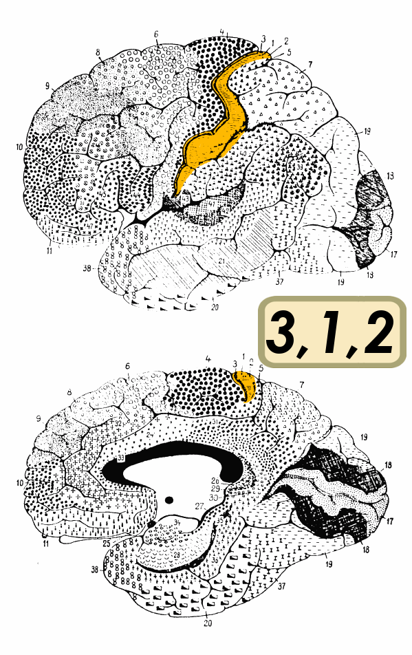
Figure 1. BA 3, 1, 2 (Diagram). Link |
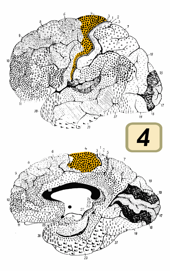
Figure 2. BA 4 (Diagram). Link |
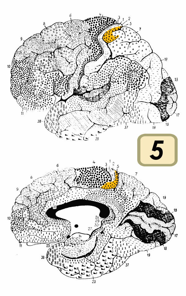
Figure 3. BA 5 (Diagram). Link |
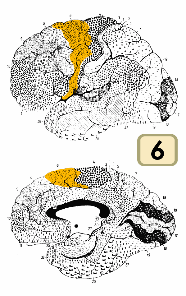
Figure 4. BA 6 (Diagram). Link |
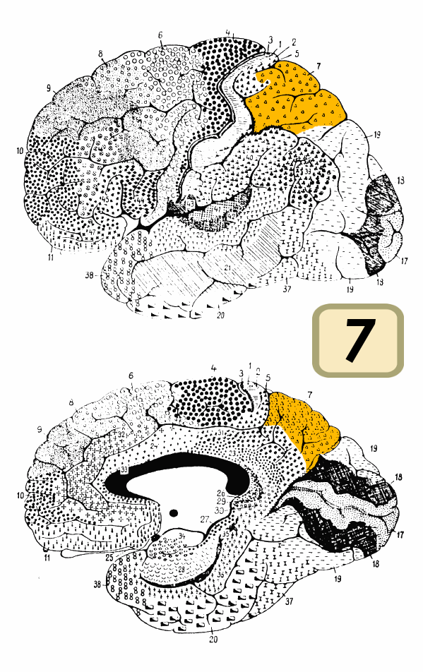
Figure 5. BA 7 (Diagram). Link |
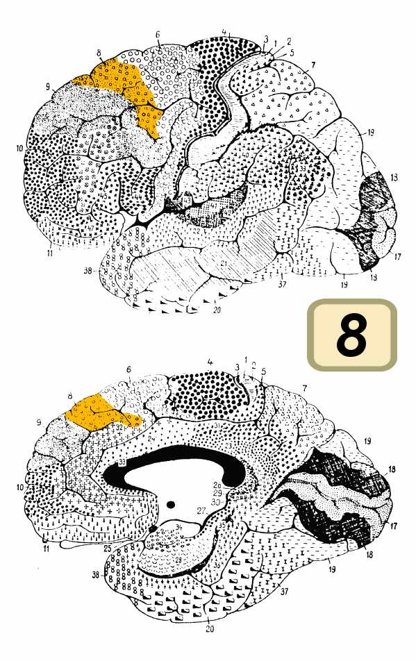
Figure 6. BA 8 (Diagram). Link |

Figure 7. BA 9 (Diagram). Link |
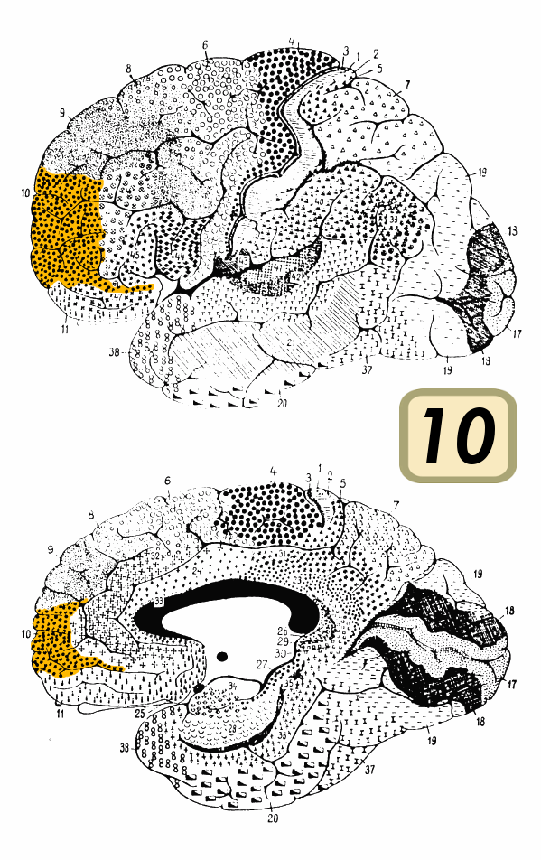
Figure 8. BA 10 (Diagram). Link |
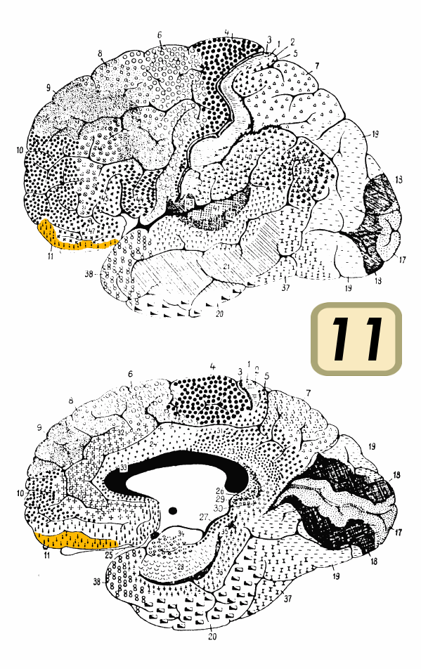
Figure 9. BA 11 (Diagram). Link |
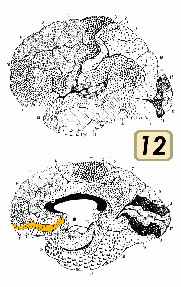
Figure 10. BA 12 (Diagram). Link |
_14.png)
Figure 11. BA 14 (Diagram). Link |
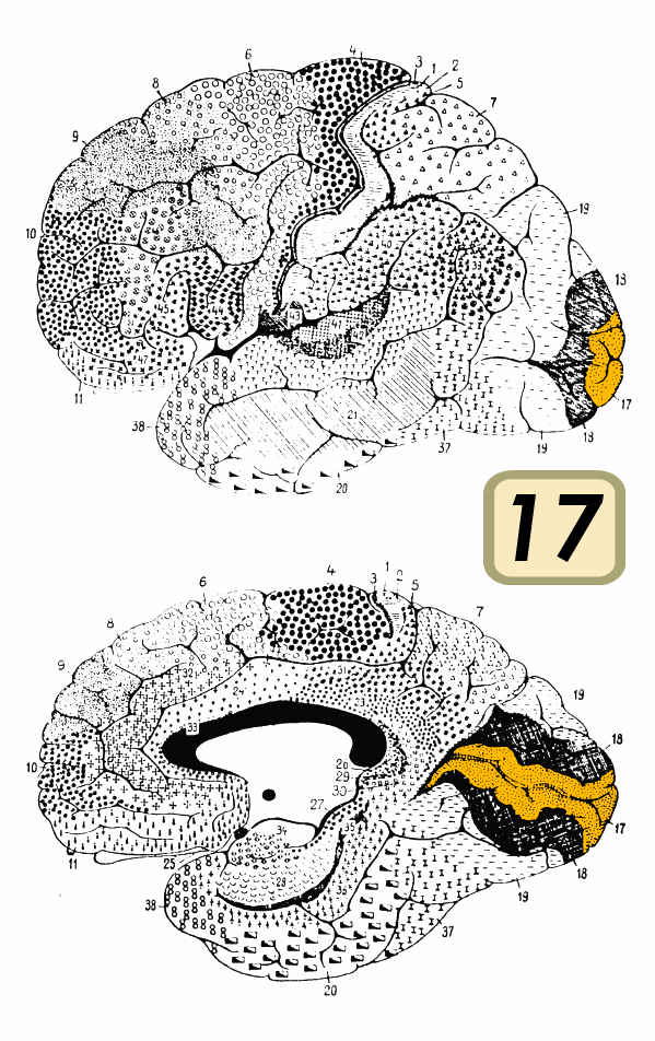
Figure 12. BA 17 (Diagram). Link |
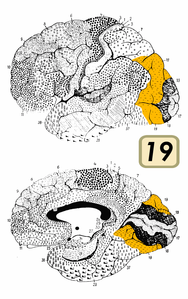
Figure 13. BA 19 (Diagram). Link |
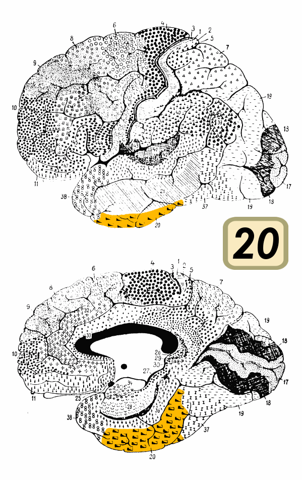
Figure 14. BA 20 (Diagram). Link |
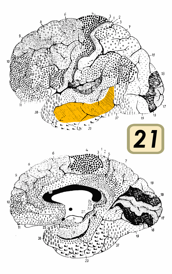
Figure 15. BA 21 (Diagram). Link |
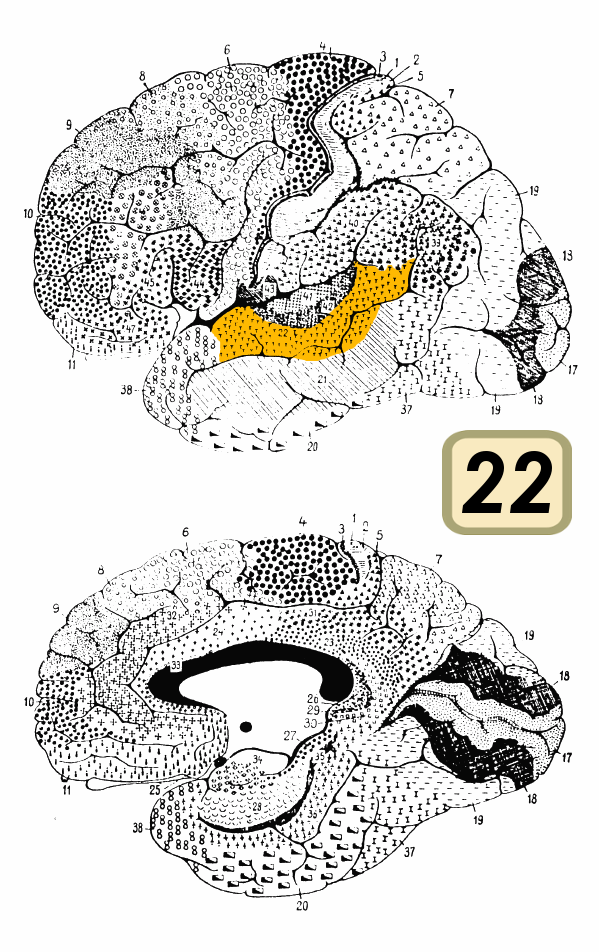
Figure 16. BA 22 (Diagram). Link |
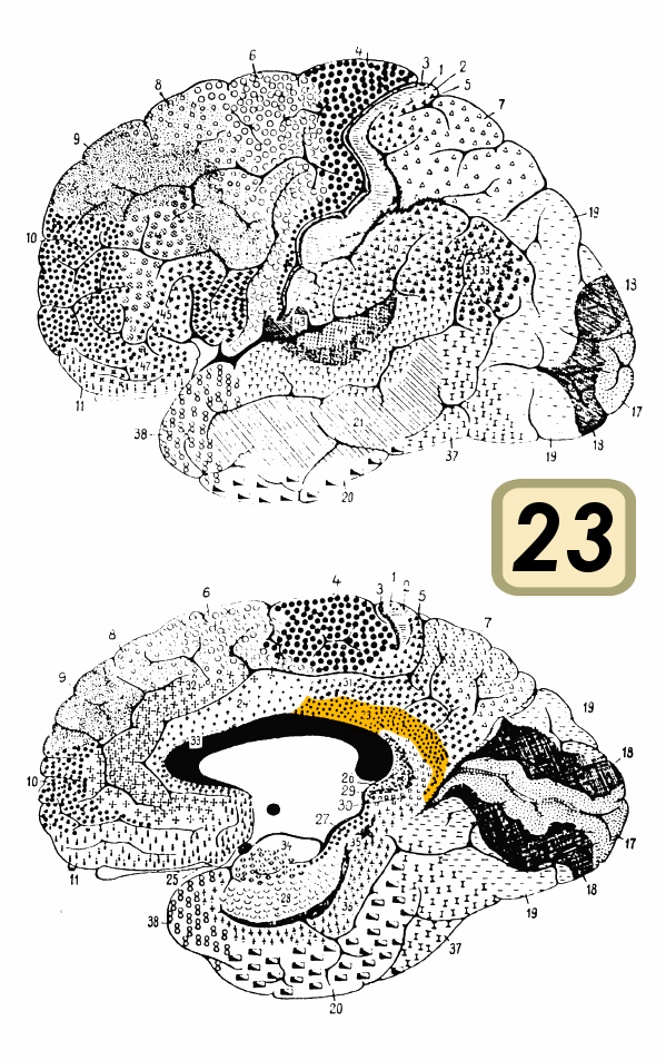
Figure 17. BA 23 (Diagram). Link |
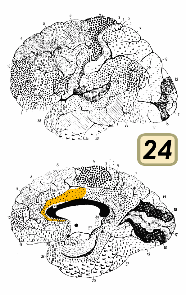
Figure 18. BA 24 (Diagram). Link |
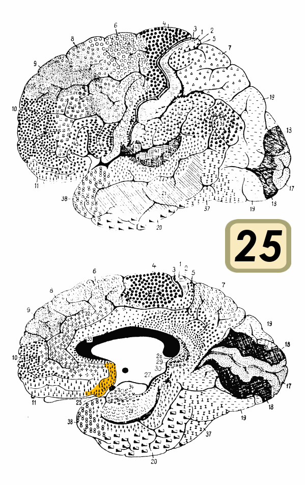
Figure 19. BA 25 (Diagram). Link |
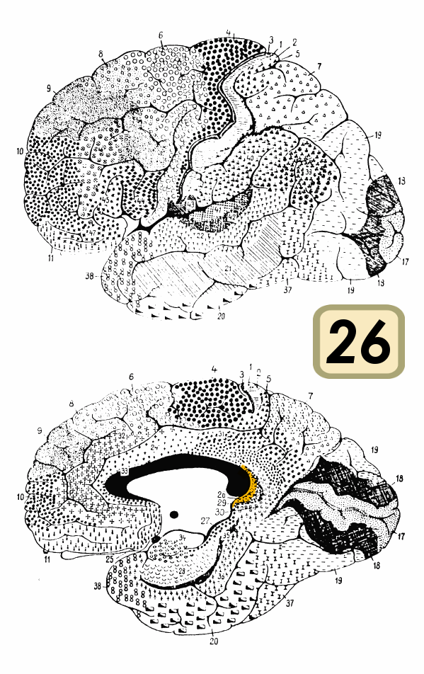
Figure 20. BA 26 (Diagram). Link |
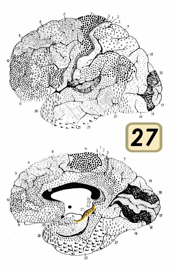
Figure 21. BA 27 (Diagram). Link |
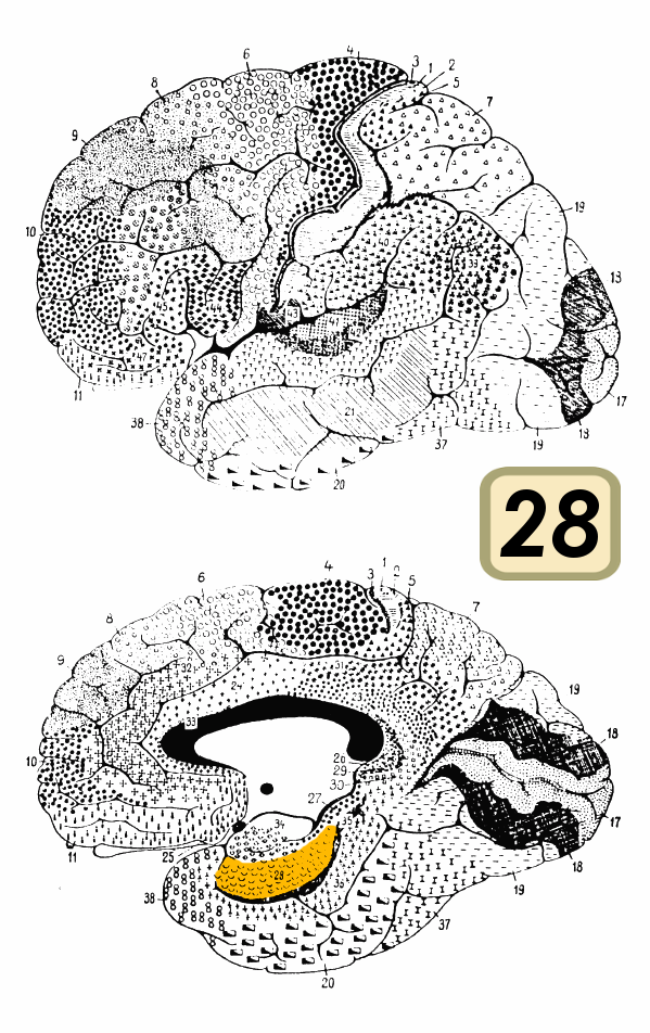
Figure 22. BA 28 (Diagram). Link |
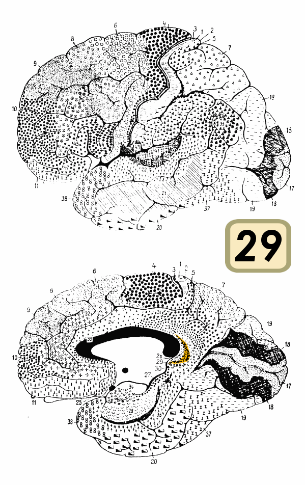
Figure 23. BA 29 (Diagram). Link |
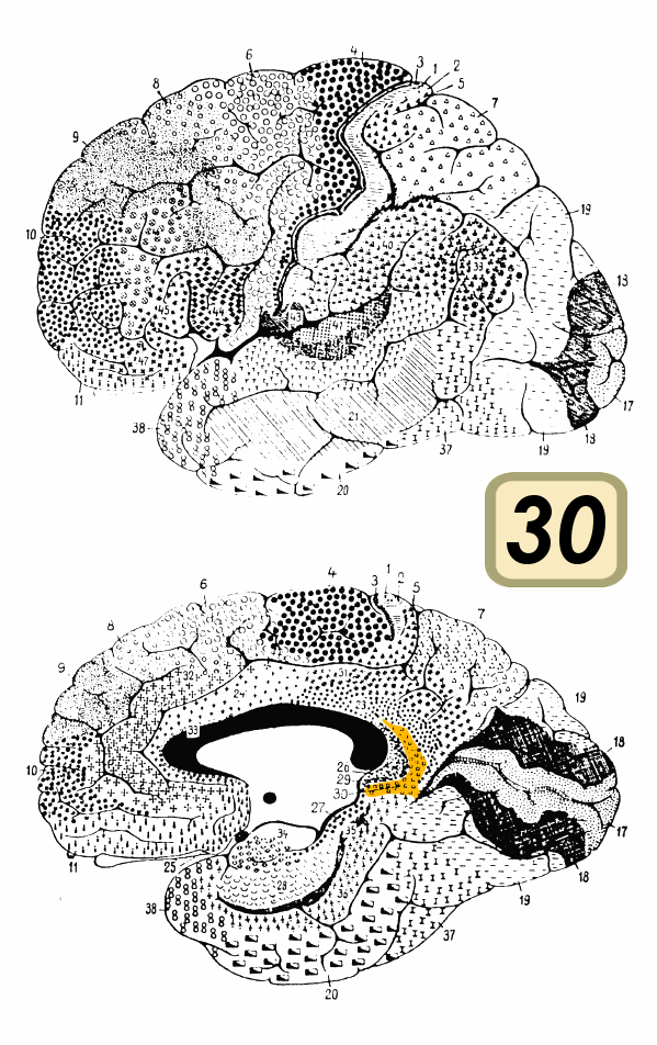
Figure 24. BA 30 (Diagram). Link |
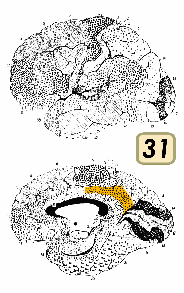
Figure 25. BA 31 (Diagram). Link |
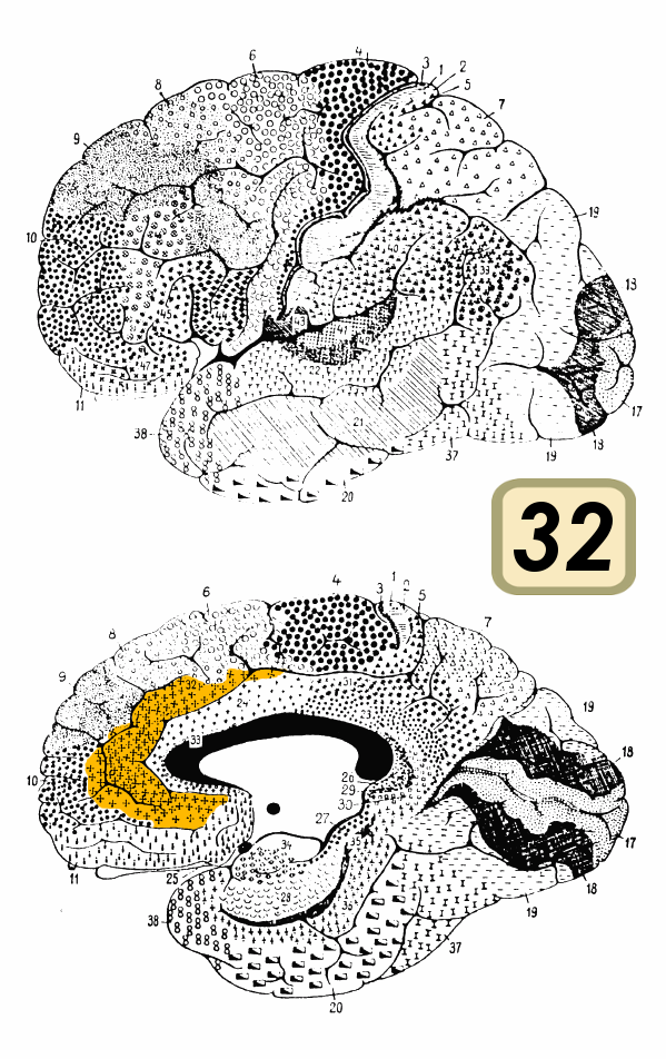
Figure 26. BA 32 (Diagram). Link |
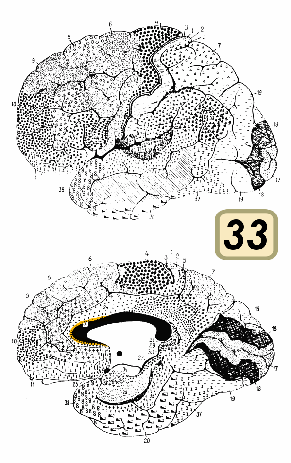
Figure 27. BA 33 (Diagram). Link |
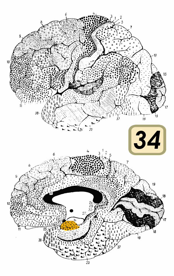
Figure 28. BA 34 (Diagram). Link |
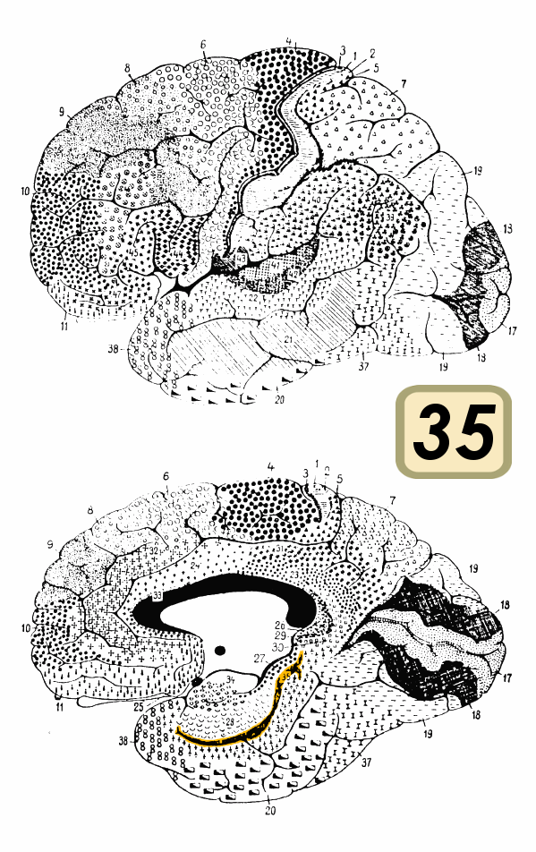
Figure 29. BA 35 (Diagram). Link |

Figure 30. BA 36 (Diagram). Link |
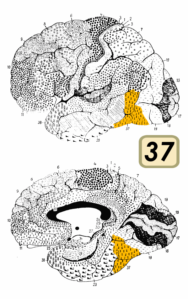
Figure 31. BA 37 (Diagram). Link |
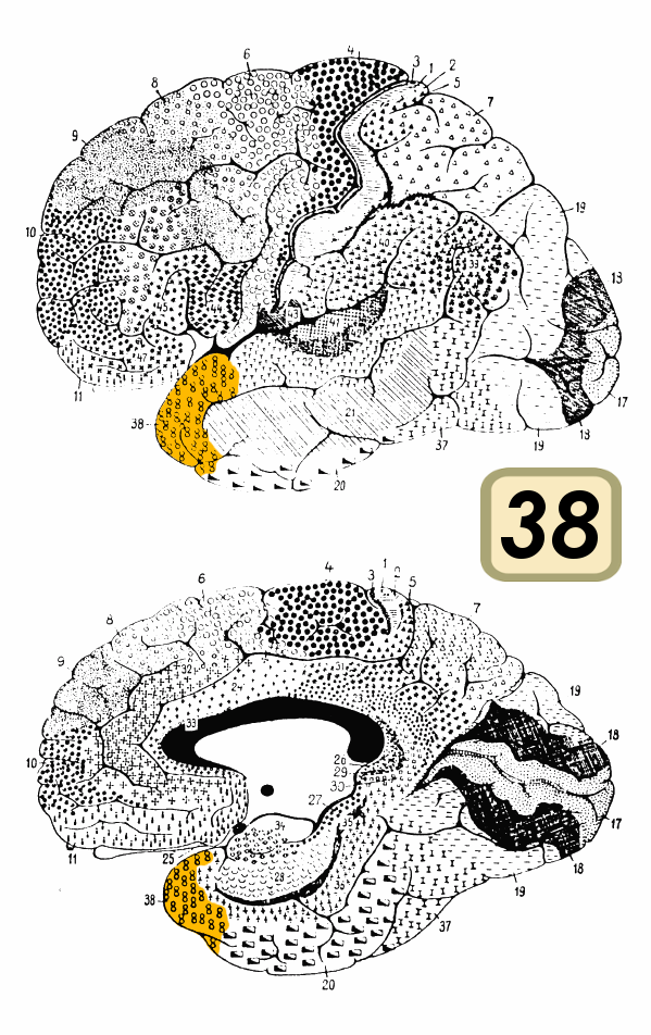
Figure 32. BA 38 (Diagram). Link |
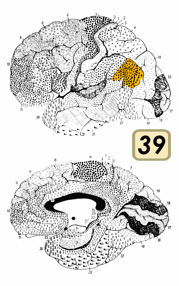
Figure 33. BA 39 (Diagram). Link |
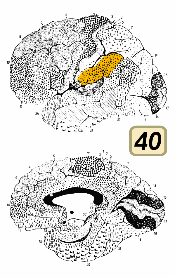
Figure 34. BA 40 (Diagram). Link |
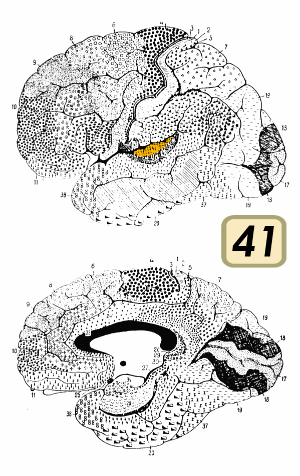
Figure 35. BA 41 (Diagram). Link |
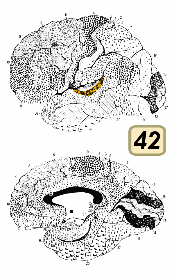
Figure 36. BA 42 (Diagram). Link |
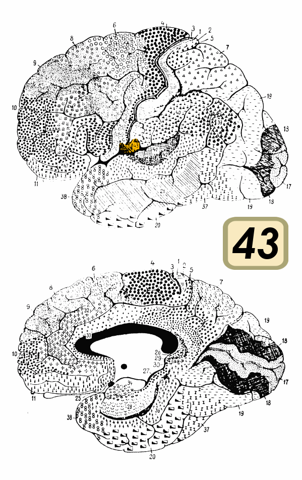
Figure 37. BA 43 (Diagram). Link |
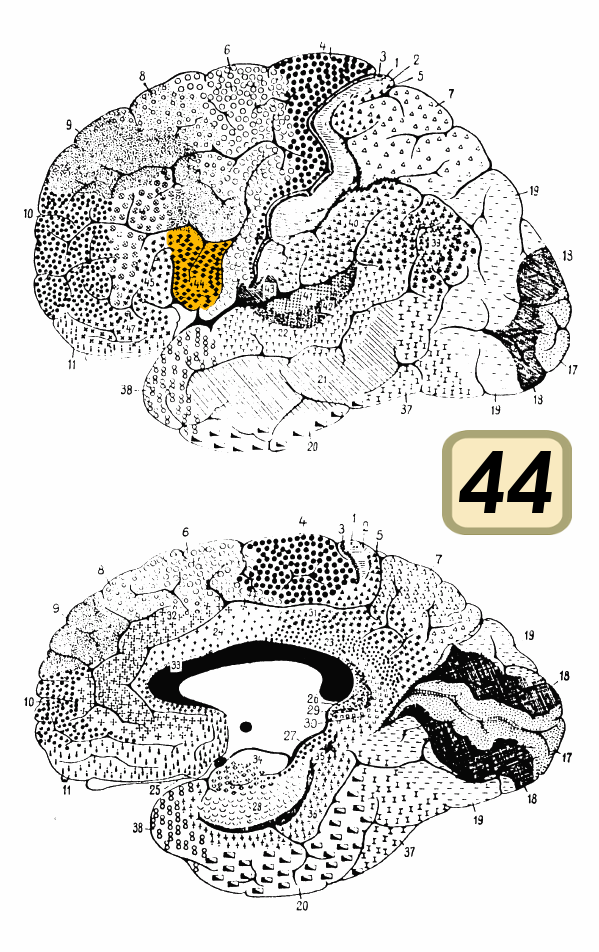
Figure 38. BA 44 (Diagram). Link |
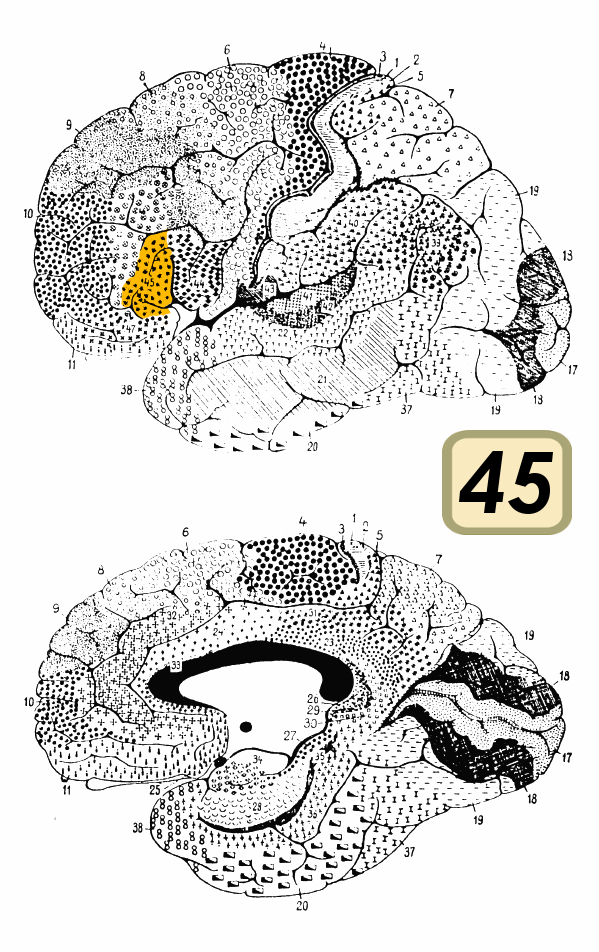
Figure 39. BA 45 (Diagram). Link |
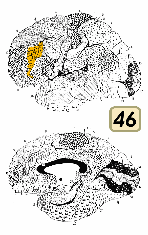
Figure 40. BA 46 (Diagram). Link |
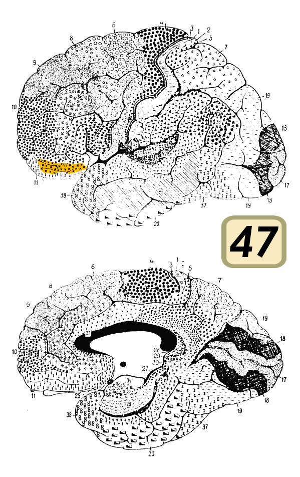
Figure 41. BA 47 (Diagram). Link |
Vesalius A. (1543) - Willis T. (1667) - Galvani L. (1791) - Bourgery J.M. (1830) - Golgi C. (1873) - Broca P. (1877) - Waldeyer (1891) - Cajal S.R. (1917) - Gray H. (1918) - Terminologia Anatomica (TA)
Andreœ Vesalii Bruxellensis scholœ medicorum Patauinœ professons, Suorum de Humani corporis fabrica librorum Epitome. Cum Cœsareœ Maiest. Galliarum Regis, ac Senatus Veneti gratia & priuilegio, ut in diplomatis eorundem continetur, Basileae : ex officina Ioannis Oporini, 1543.
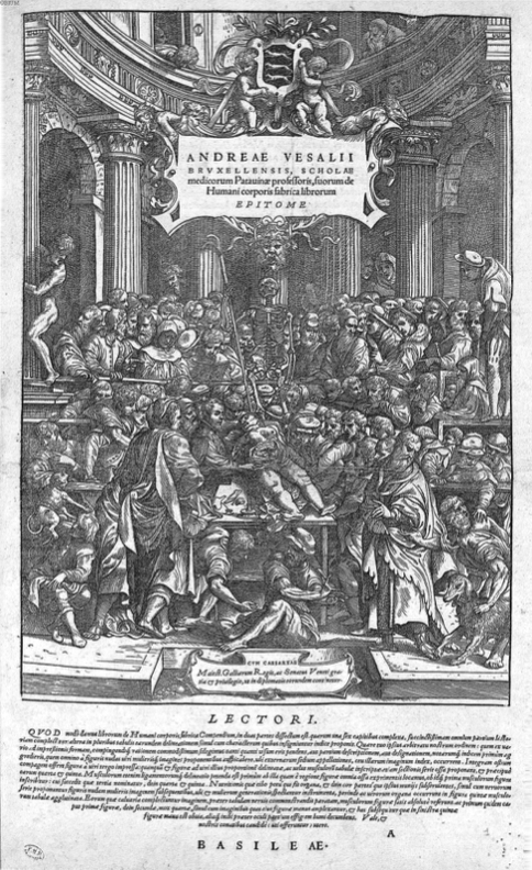
Figure 1. Vesalius, A., Suorum de Humani corporis fabrica librorum Epitome (p. 1), 1543. Link |
Figure 2. Vesalius, A., Suorum de Humani corporis fabrica librorum Epitome (p. 2), 1543. Link |
Figure 3. Vesalius, A., Suorum de Humani corporis fabrica librorum Epitome (p. 13), 1543. Link |
Figure 4. Vesalius, A., Suorum de Humani corporis fabrica librorum Epitome (p. 14), 1543. Link |
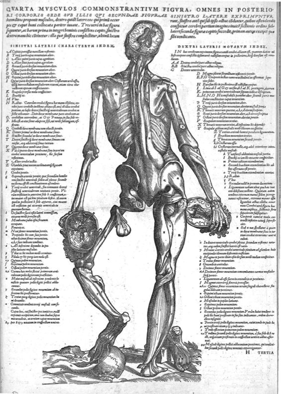
Figure 1. Vesalius, A., Suorum de Humani corporis fabrica librorum Epitome (p. 15), 1543. Link |
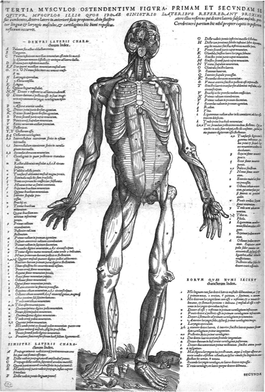
Figure 2. Vesalius, A., Suorum de Humani corporis fabrica librorum Epitome (p. 16), 1543. Link |
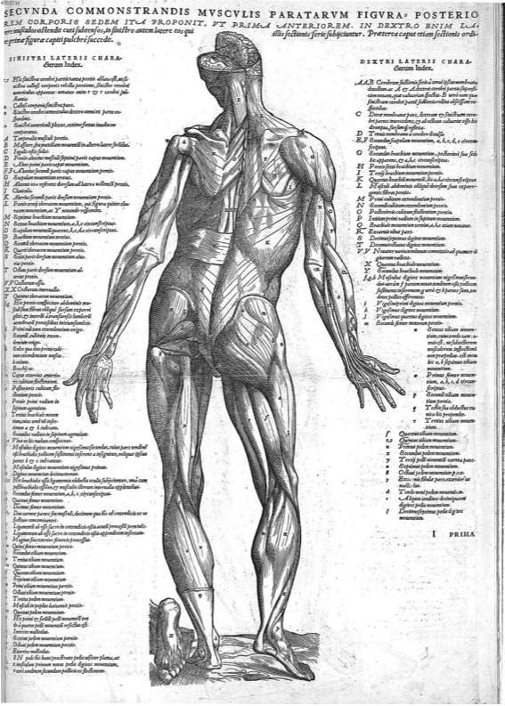
Figure 3. Vesalius, A., Suorum de Humani corporis fabrica librorum Epitome (p. 17), 1543. Link |
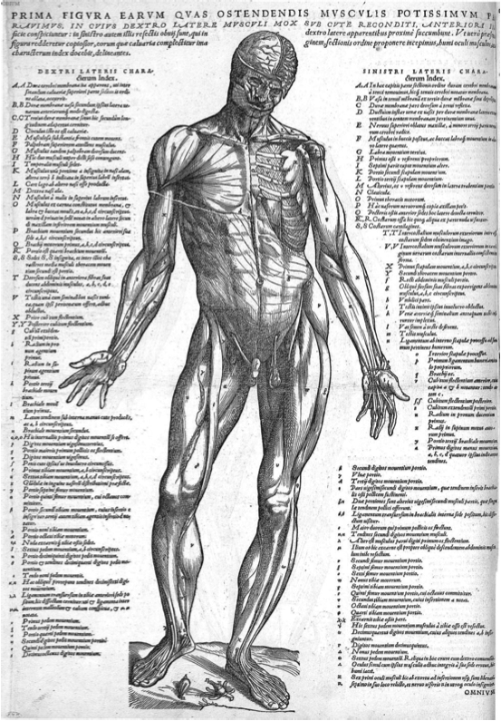
Figure 4. Vesalius, A., Suorum de Humani corporis fabrica librorum Epitome (p. 18), 1543. Link |
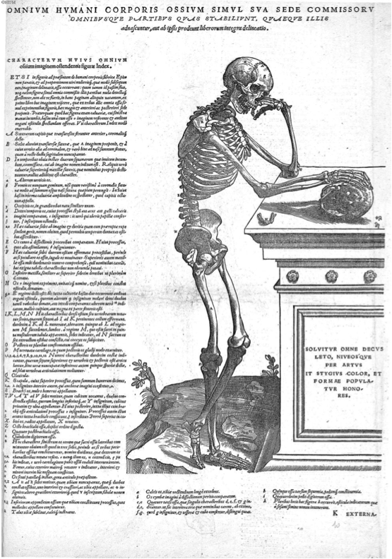
Figure 5. Vesalius, A., Suorum de Humani corporis fabrica librorum Epitome (p. 19), 1543. Link |
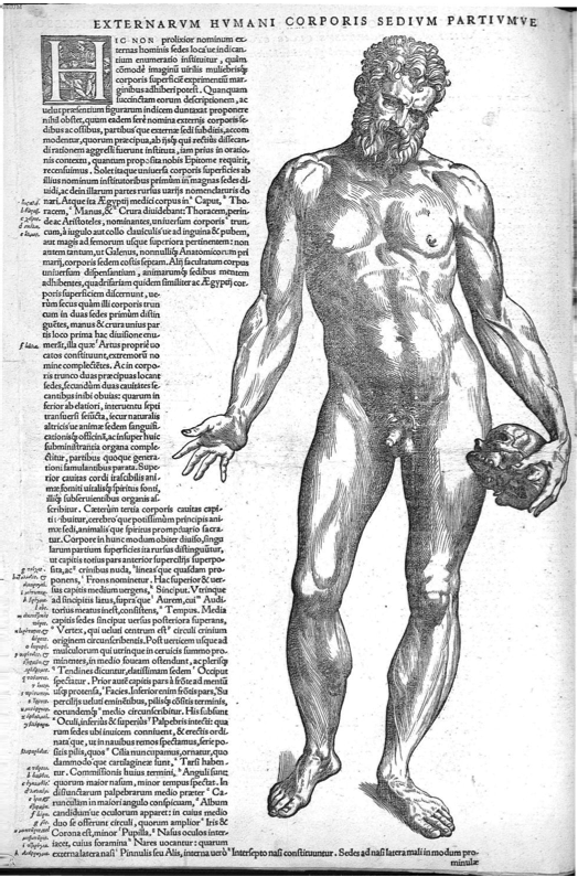
Figure 6. Vesalius, A., Suorum de Humani corporis fabrica librorum Epitome (p. 20), 1543. Link |
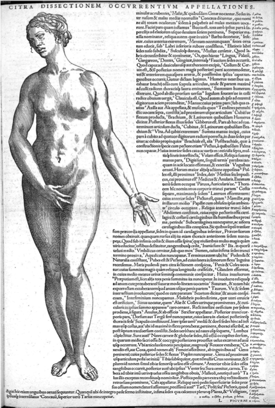
Figure 7. Vesalius, A., Suorum de Humani corporis fabrica librorum Epitome (p. 21), 1543. Link |
Figure 8. Vesalius, A., Suorum de Humani corporis fabrica librorum Epitome (p. 22), 1543. Link |
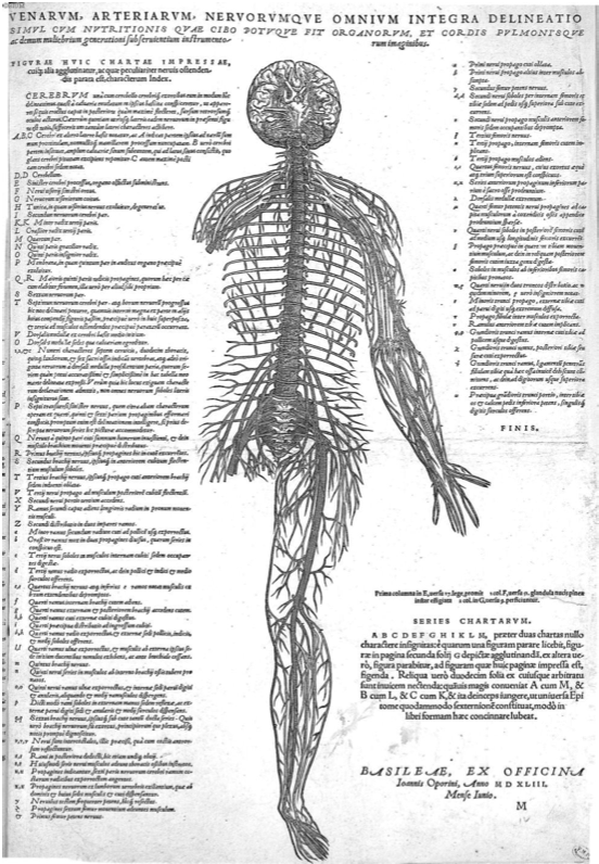
Figure 9. Vesalius, A., Suorum de Humani corporis fabrica librorum Epitome (p. 23), 1543. Link |
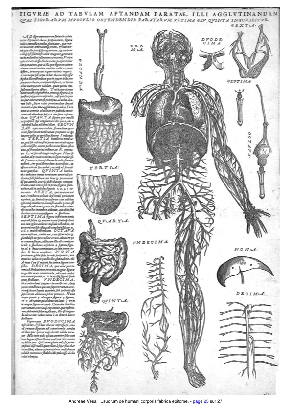
Figure 10. Vesalius, A., Suorum de Humani corporis fabrica librorum Epitome (p. 25), 1543. Link |
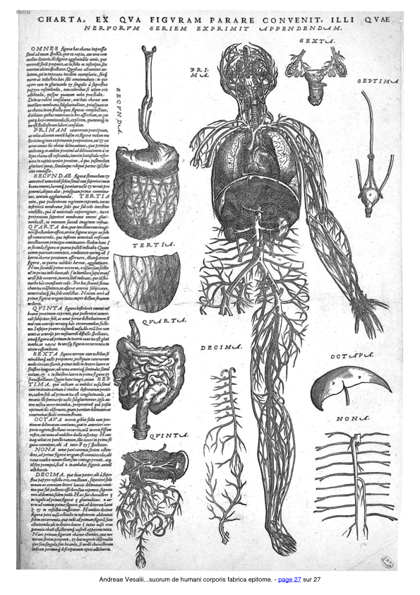
Figure 11. Vesalius, A., Suorum de Humani corporis fabrica librorum Epitome (p. 27), 1543. Link |
Andreœ Vesalii Bruxellensis, Scholœ medicorum Paîauinœ professons, de Humani corporis fabrica Libri septem, cum Cœsarœ Maiest. Galliarum Regis, ac Senatus Veneti gratia & priui- legio, ut in diplomatis eorundem continetur, Basileae : ex officina Ioannis Oporini, 1543.
Introduction
Book 1: The Bones and Cartilages
Book 2: The Ligaments and Muscles
Book 3: The Veins and Arteries
Book 4: The Nerves
Book 5: The Organs of Nutrition and Generation
Book 6: The Heart and Associated Organs
Book 7: The Brain
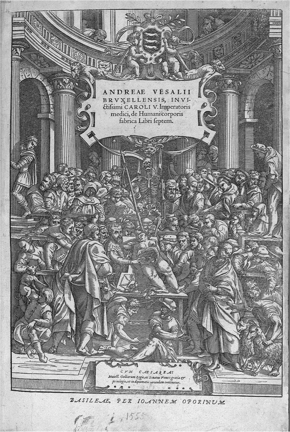
Figure 1. Vesalius, A., de Humani corporis fabrica Libri septem (page de couverture), 1543. Link |
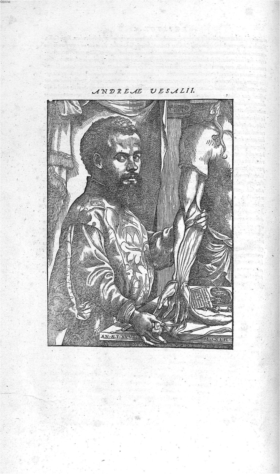
Figure 2. Vesalius, A., de Humani corporis fabrica Libri septem (p. i), 1543. Link |
Book 1: The Bones and Cartilages
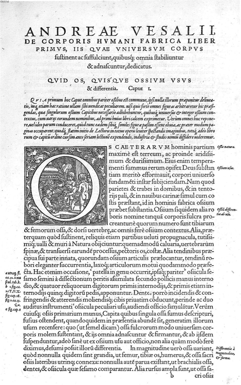
Figure 1. Vesalius, A., de Humani corporis fabrica Libri septem (p. 1), 1543. Link |
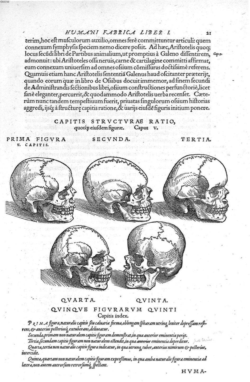
Figure 2. Vesalius, A., de Humani corporis fabrica Libri septem (p. 21), 1543. Link |
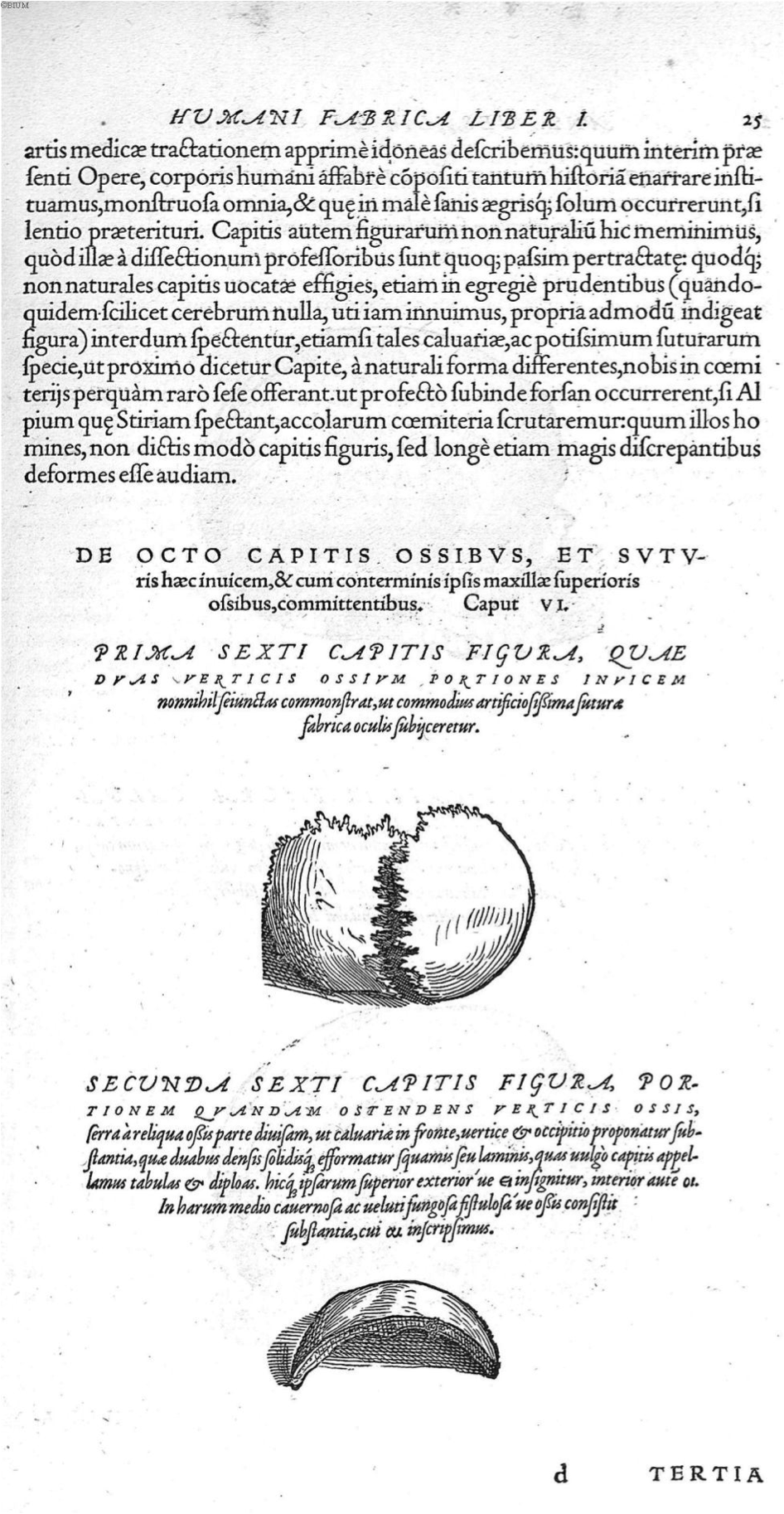
Figure 3. Vesalius, A., de Humani corporis fabrica Libri septem (p. 25), 1543. Link |
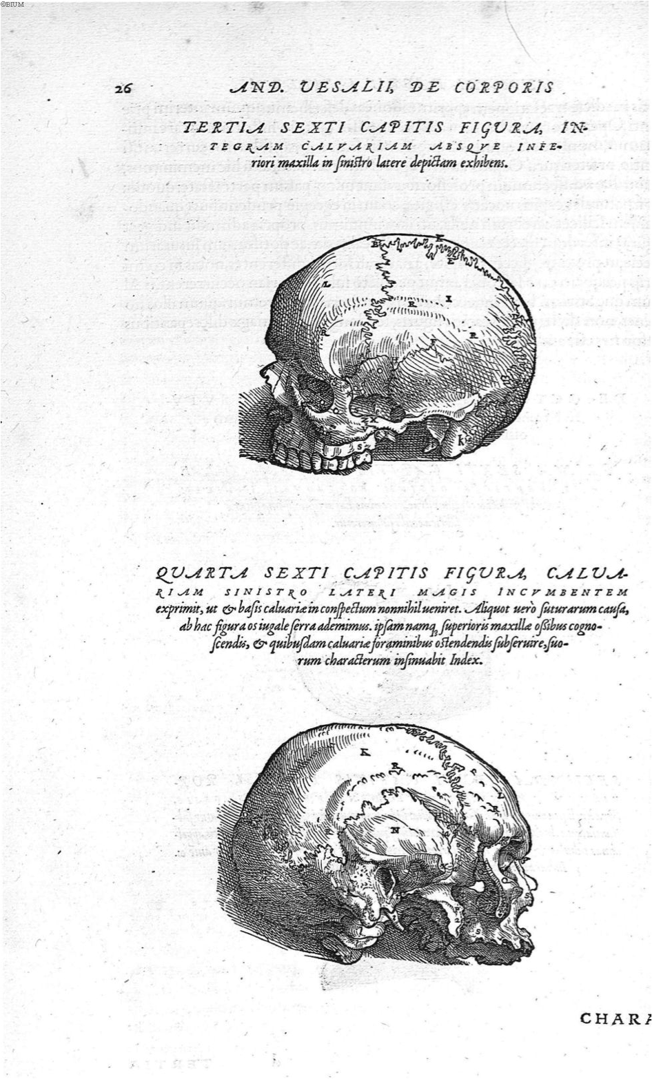
Figure 4. Vesalius, A., de Humani corporis fabrica Libri septem (p. 26), 1543. Link |
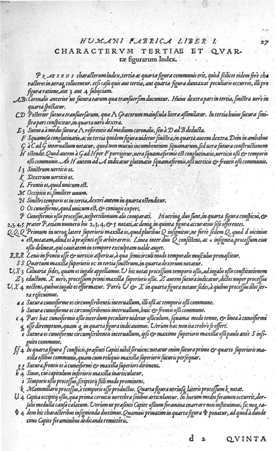
Figure 5. Vesalius, A., de Humani corporis fabrica Libri septem (p. 27), 1543. Link |
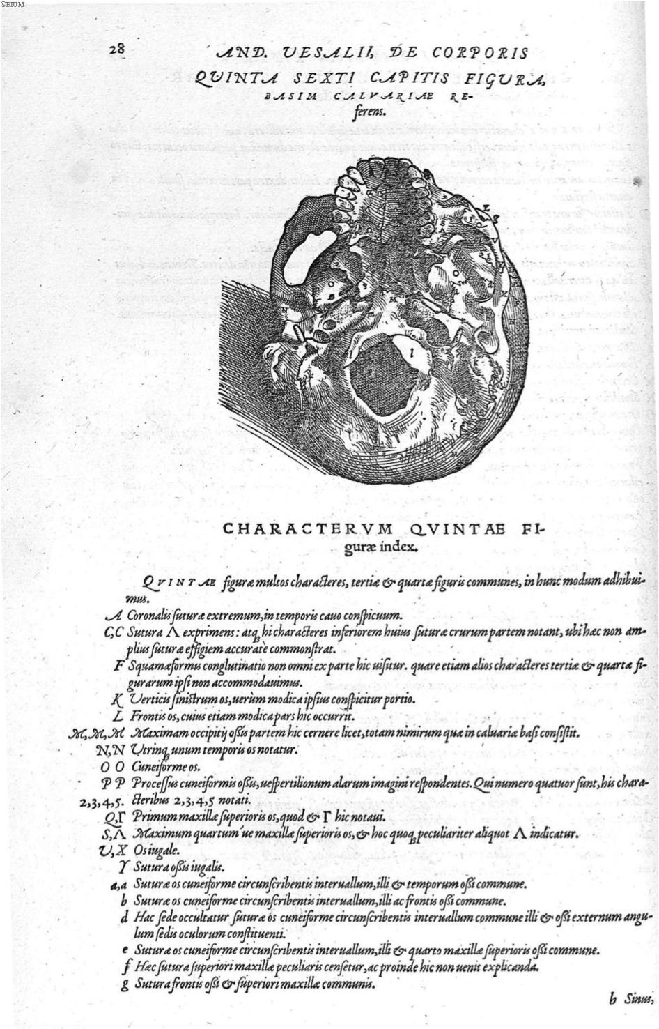
Figure 6. Vesalius, A., de Humani corporis fabrica Libri septem (p. 28), 1543. Link |
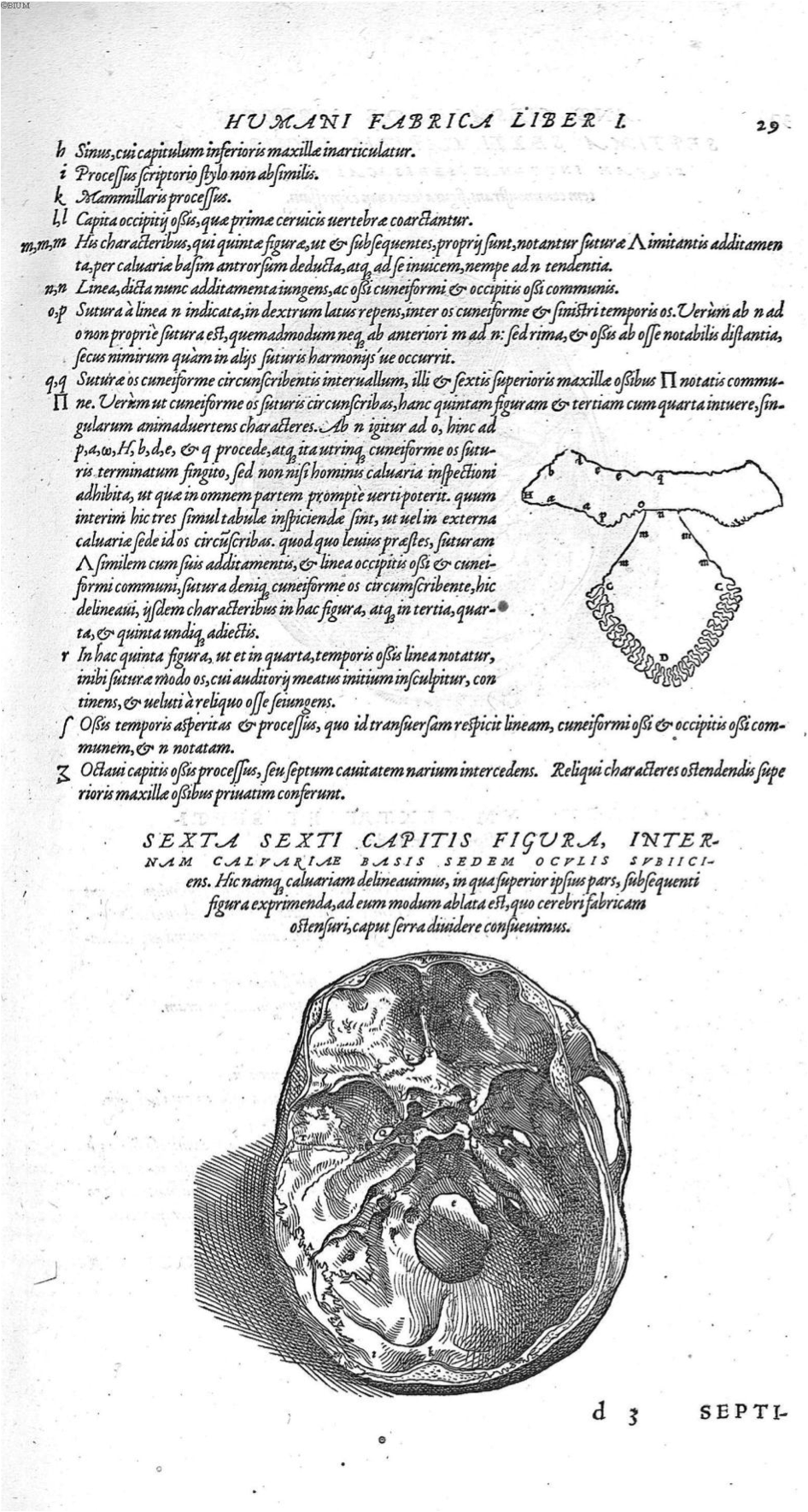
Figure 7. Vesalius, A., de Humani corporis fabrica Libri septem (p. 29), 1543. Link |
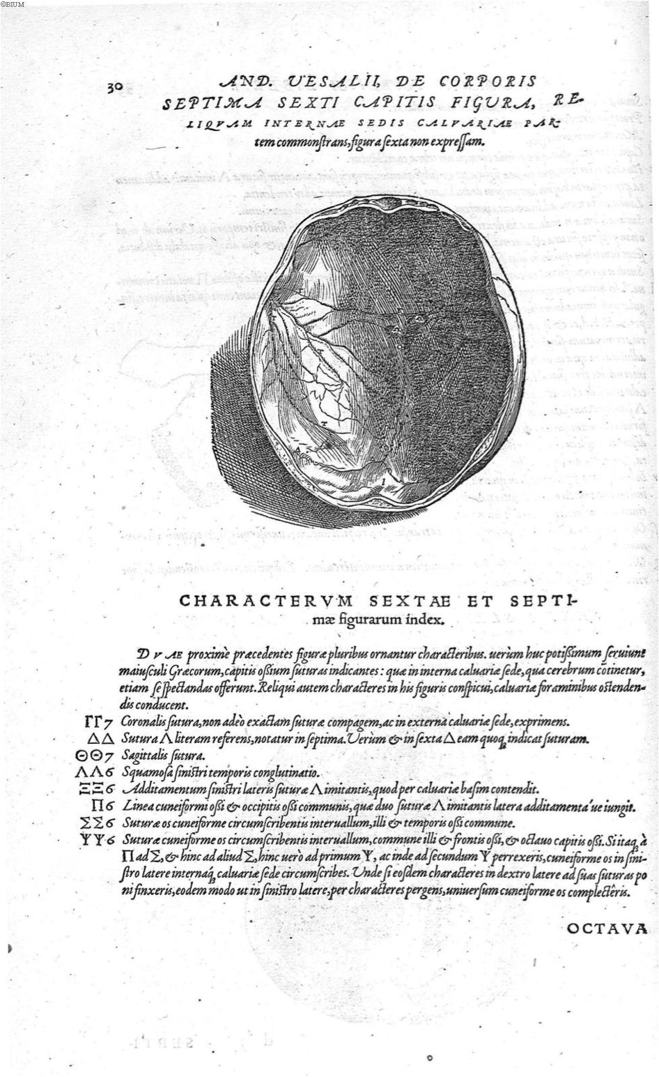
Figure 8. Vesalius, A., de Humani corporis fabrica Libri septem (p. 30), 1543. Link |
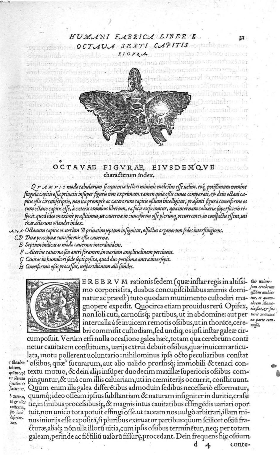
Figure 9. Vesalius, A., de Humani corporis fabrica Libri septem (p. 31), 1543. Link |
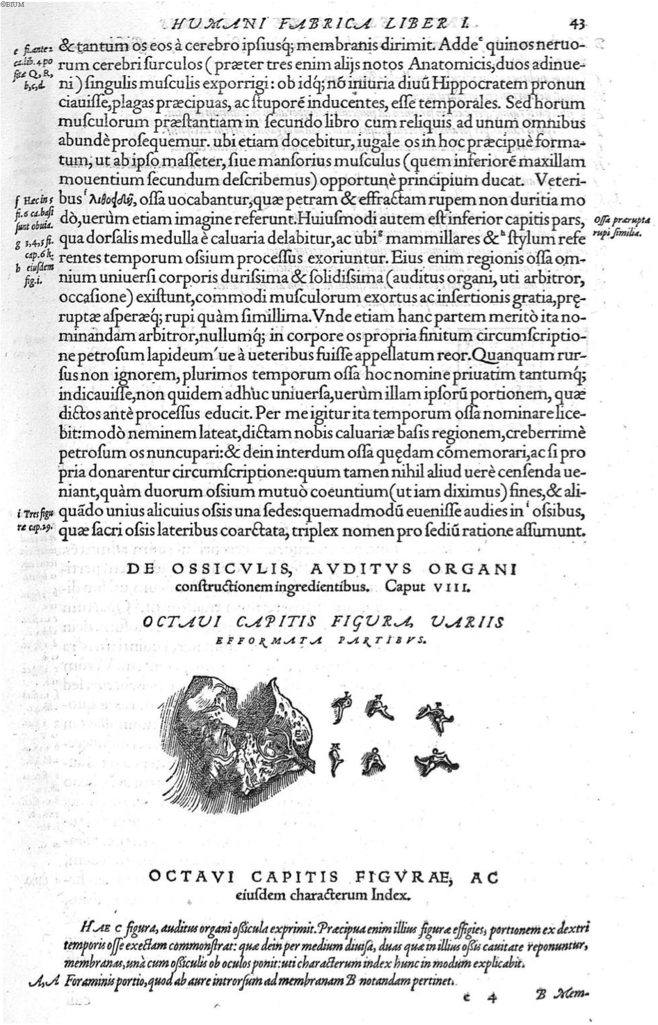
Figure 10. Vesalius, A., de Humani corporis fabrica Libri septem (p. 43), 1543. Link |
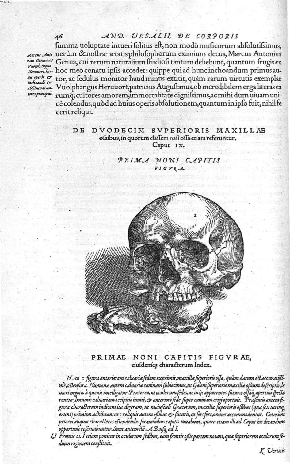
Figure 11. Vesalius, A., de Humani corporis fabrica Libri septem (p. 46), 1543. Link |
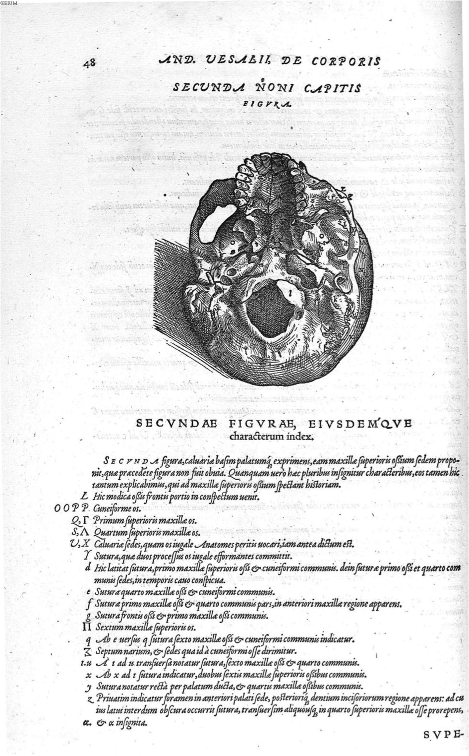
Figure 12. Vesalius, A., de Humani corporis fabrica Libri septem (p. 48), 1543. Link |
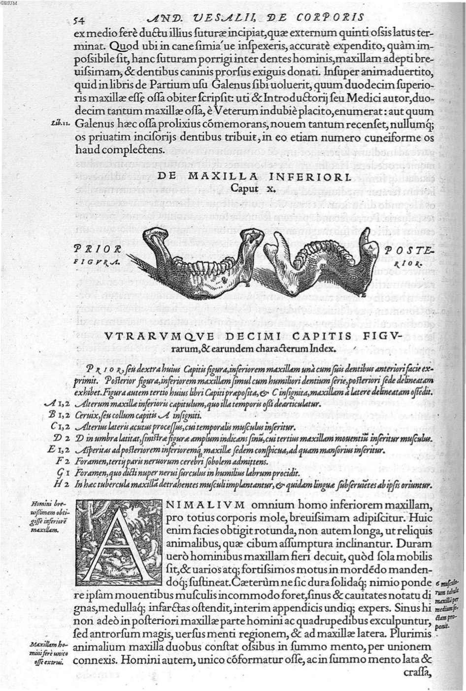
Figure 13. Vesalius, A., de Humani corporis fabrica Libri septem (p. 54), 1543. Link |
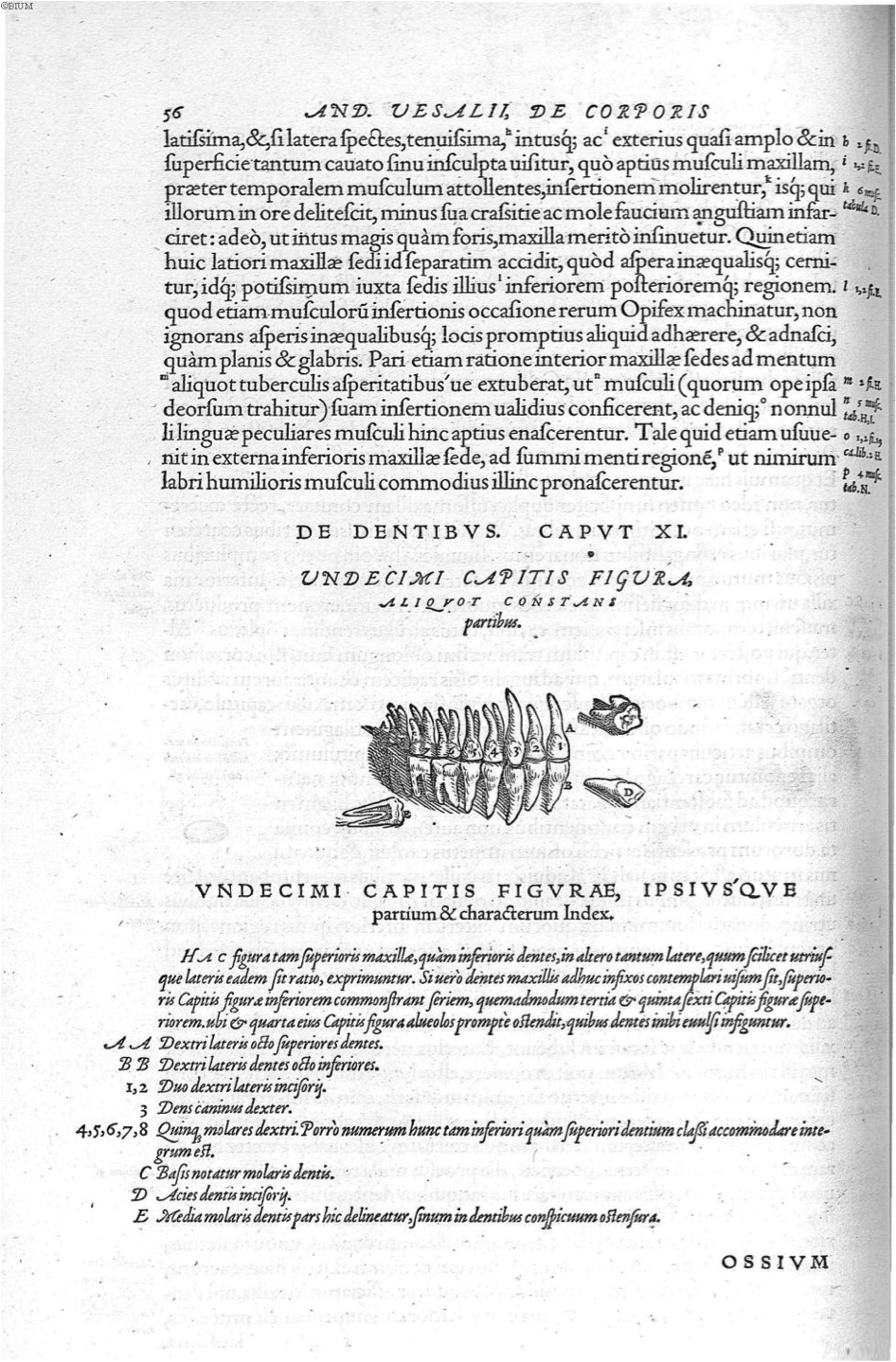
Figure 14. Vesalius, A., de Humani corporis fabrica Libri septem (p. 56), 1543. Link |
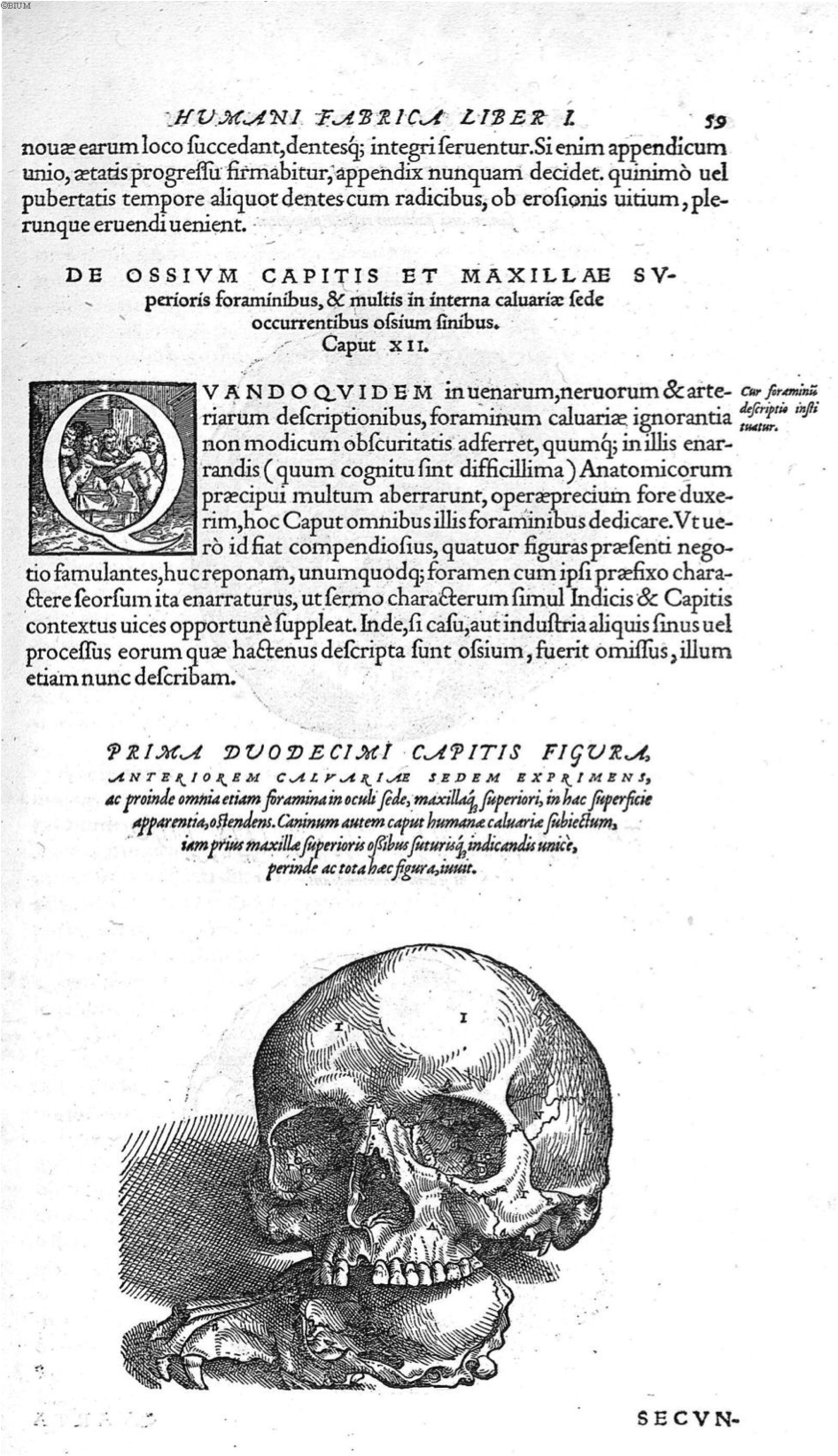
Figure 15. Vesalius, A., de Humani corporis fabrica Libri septem (p. 59), 1543. Link |
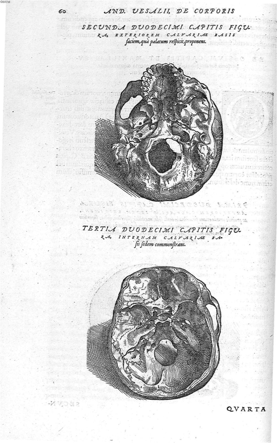
Figure 16. Vesalius, A., de Humani corporis fabrica Libri septem (p. 60), 1543. Link |
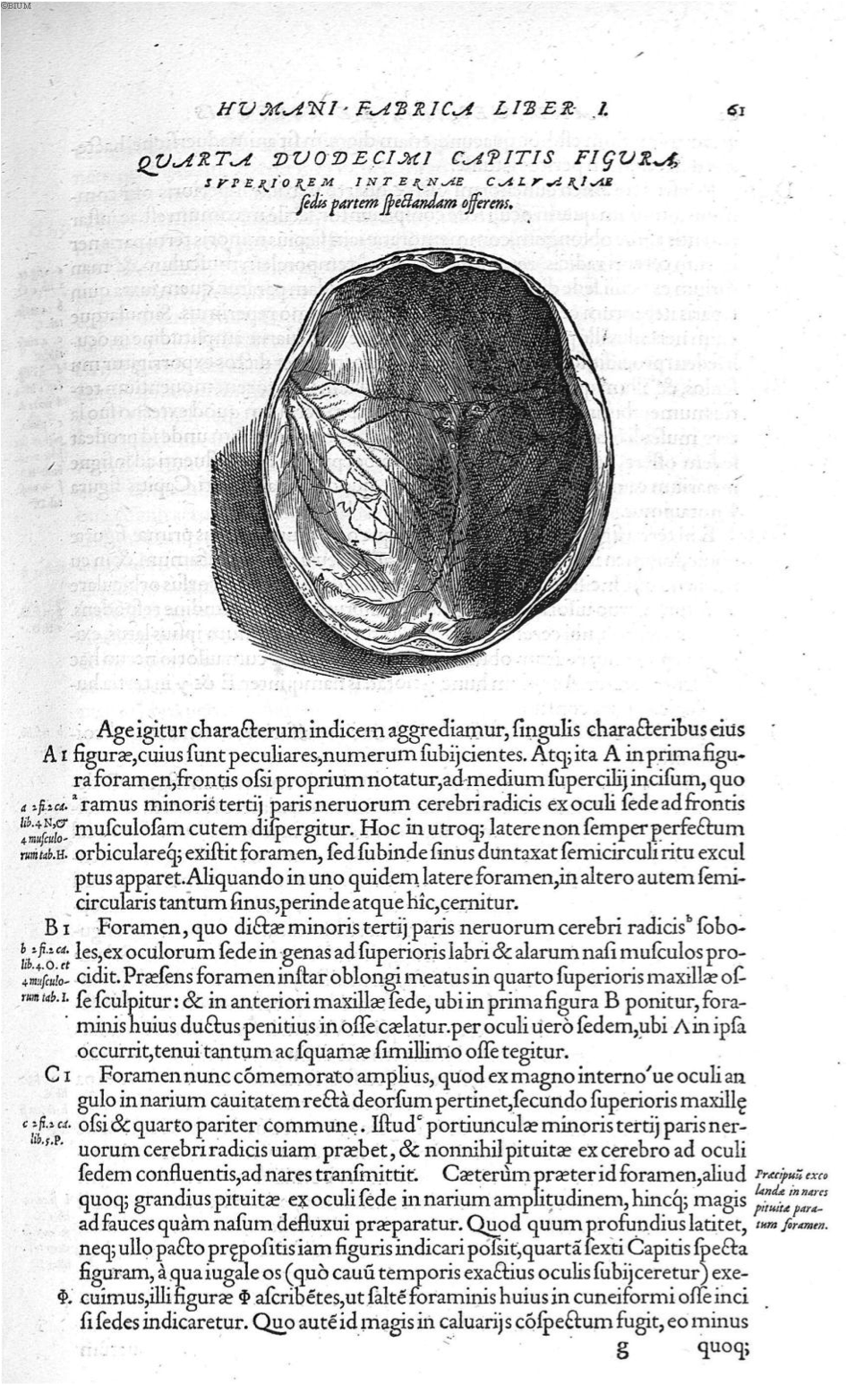
Figure 17. Vesalius, A., de Humani corporis fabrica Libri septem (p. 61), 1543. Link |
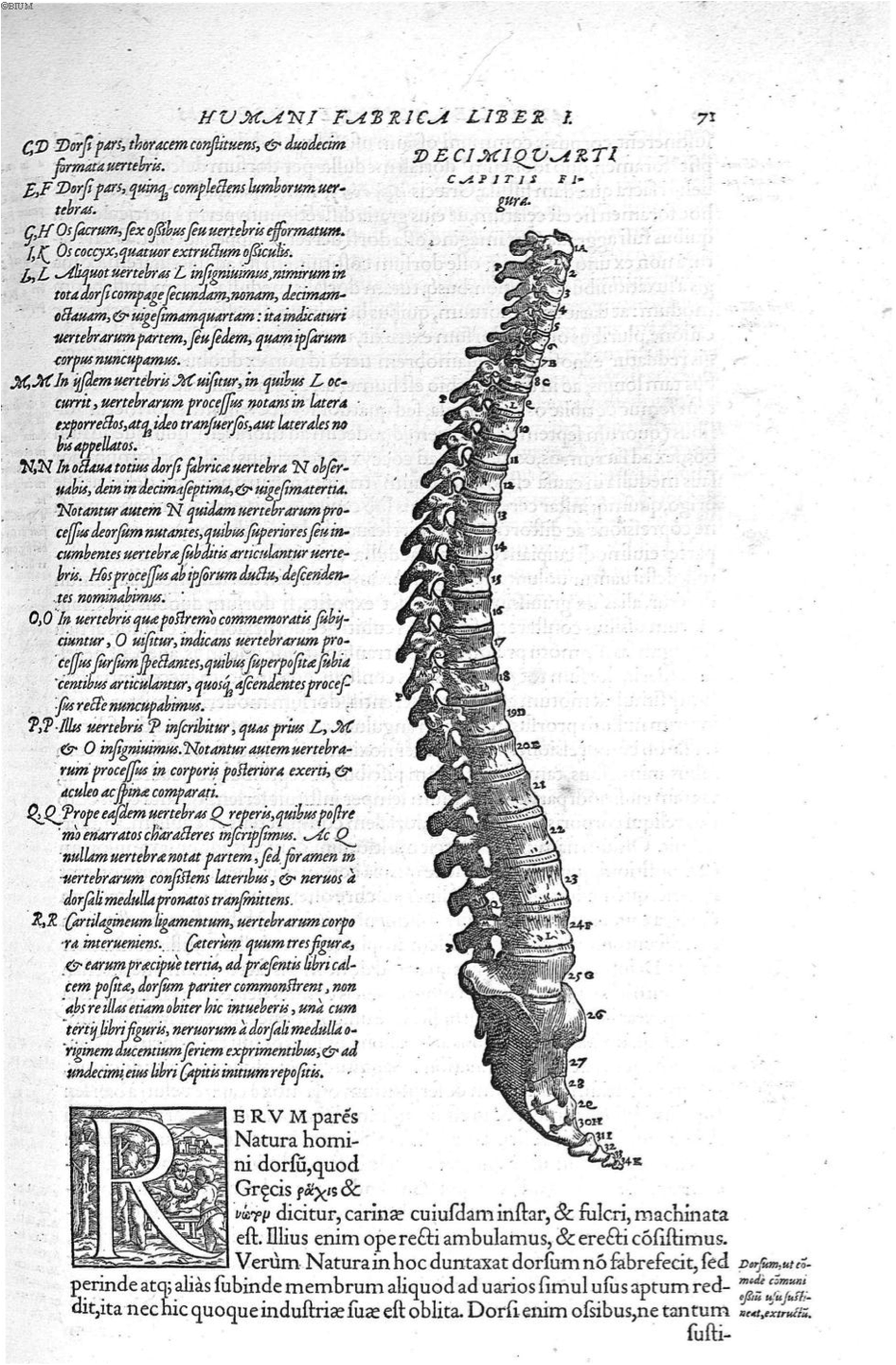
Figure 18. Vesalius, A., de Humani corporis fabrica Libri septem (p. 71), 1543. Link |
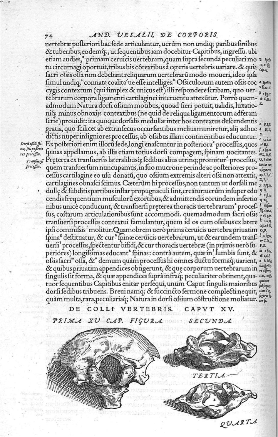
Figure 19. Vesalius, A., de Humani corporis fabrica Libri septem (p. 74), 1543. Link |
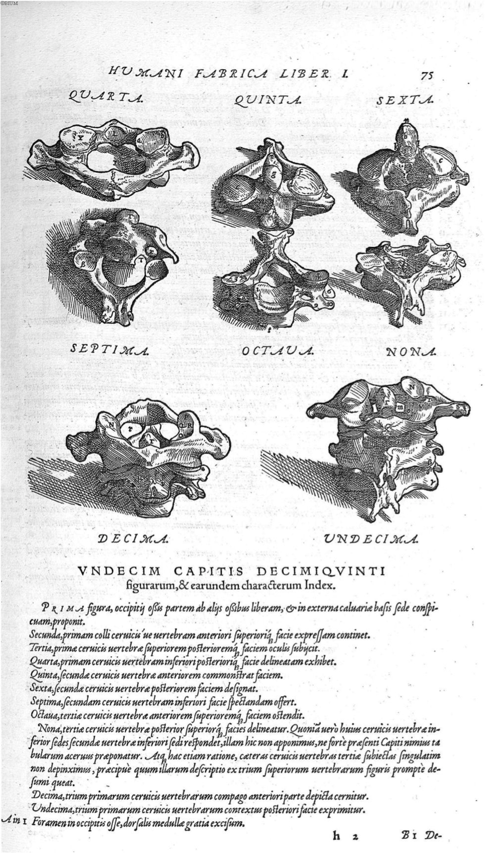
Figure 20. Vesalius, A., de Humani corporis fabrica Libri septem (p. 75), 1543. Link |
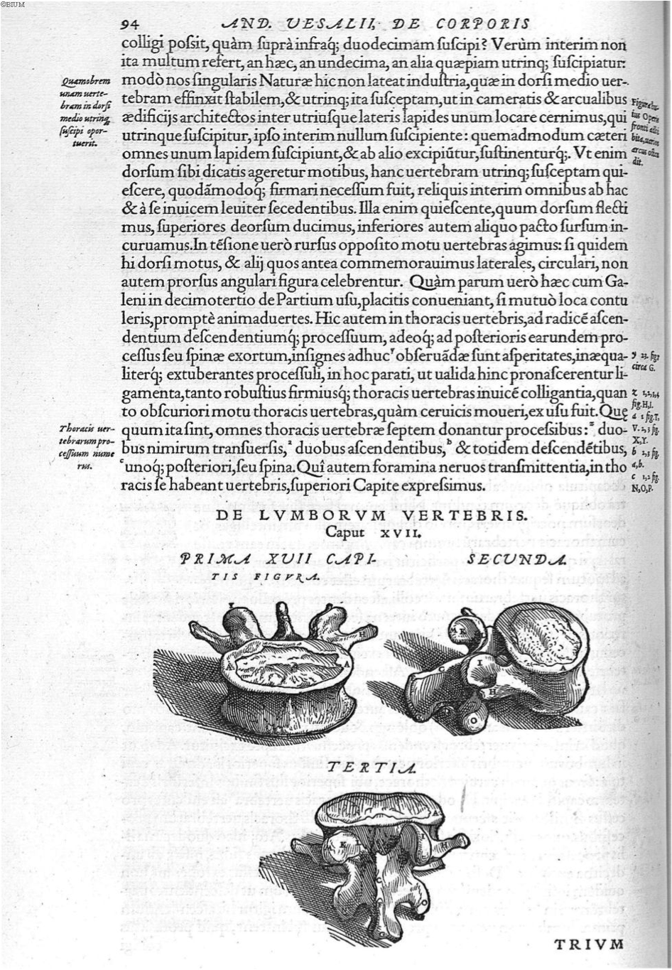
Figure 21. Vesalius, A., de Humani corporis fabrica Libri septem (p. 94), 1543. Link |
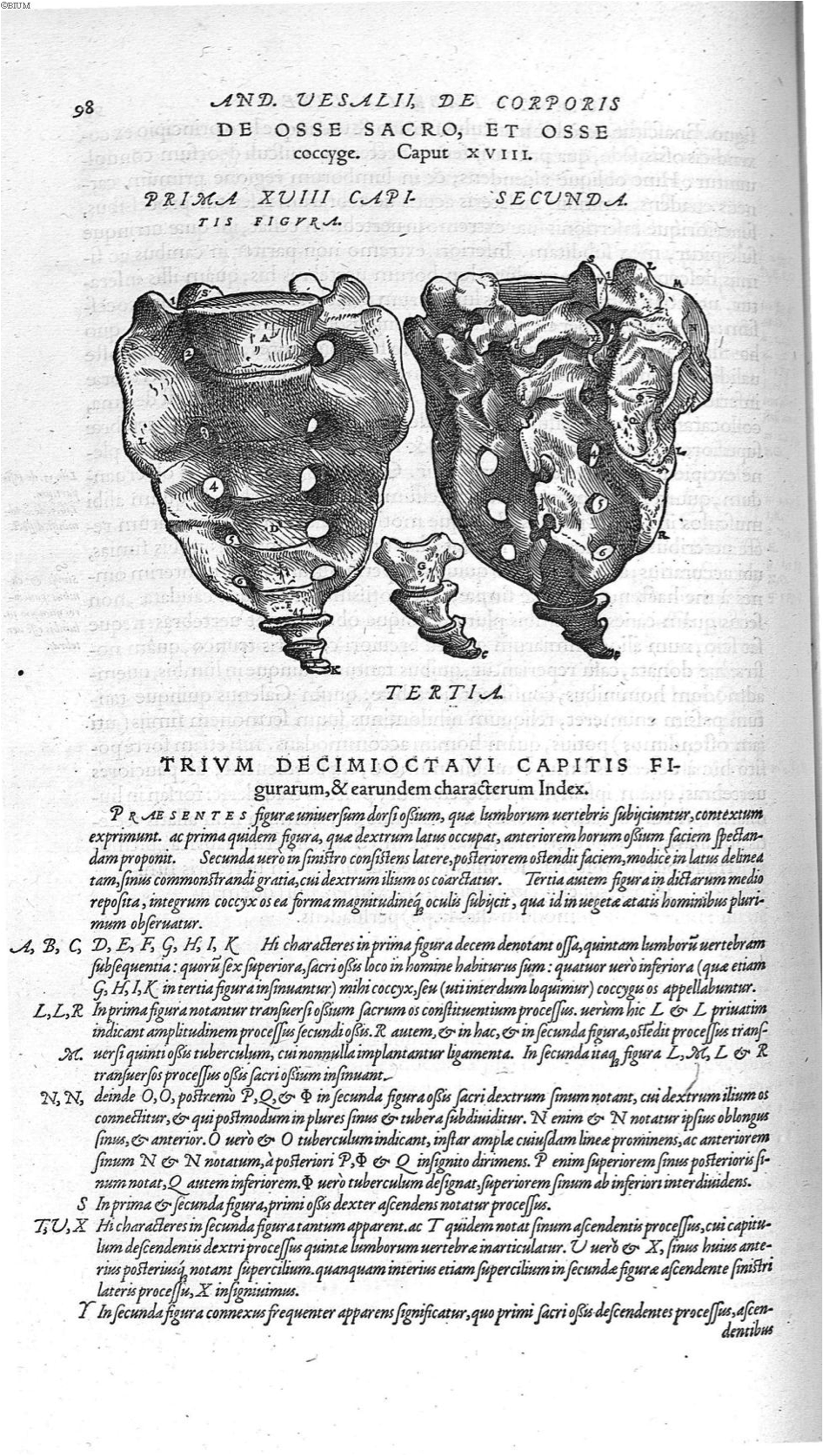
Figure 22. Vesalius, A., de Humani corporis fabrica Libri septem (p. 98), 1543. Link |
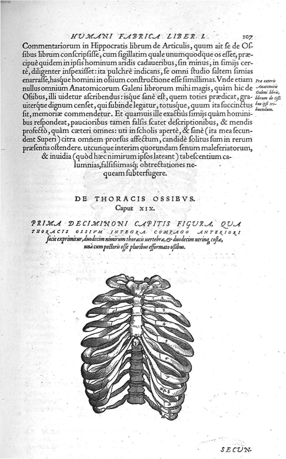
Figure 23. Vesalius, A., de Humani corporis fabrica Libri septem (p. 107), 1543. Link |
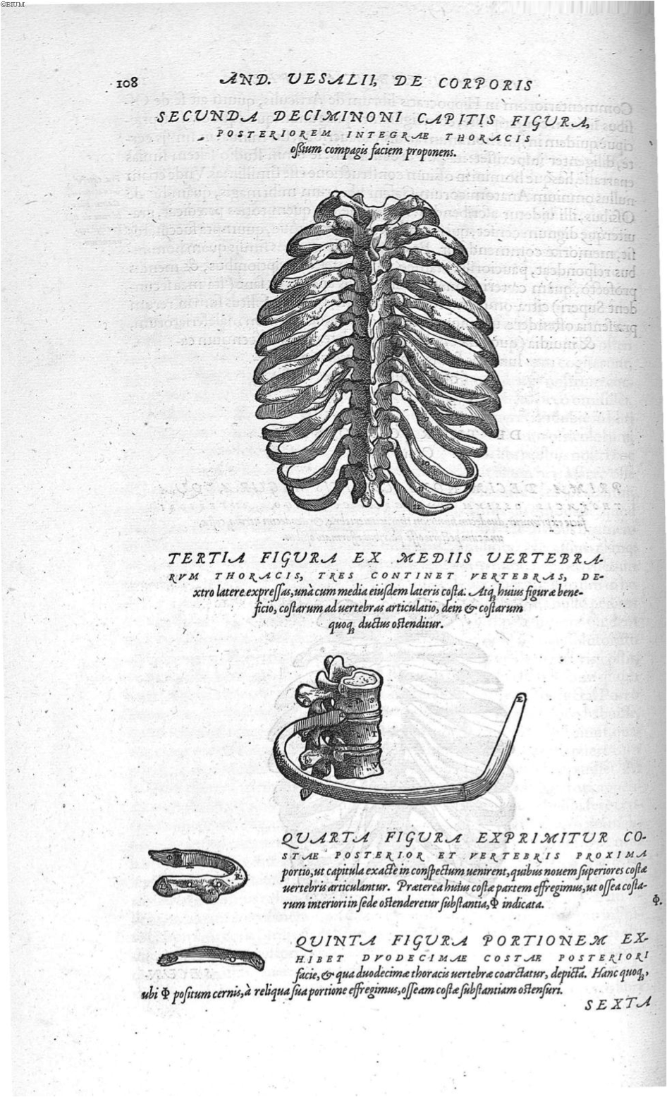
Figure 24. Vesalius, A., de Humani corporis fabrica Libri septem (p. 108), 1543. Link |
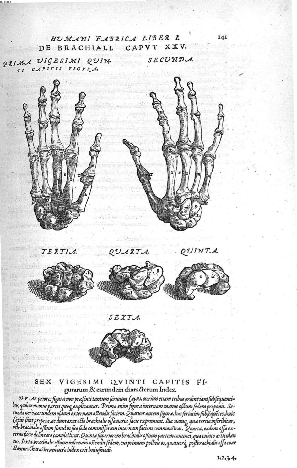
Figure 25. Vesalius, A., de Humani corporis fabrica Libri septem (p. 141), 1543. Link |
Figure 26. Vesalius, A., de Humani corporis fabrica Libri septem (p. 184), 1543. Link |
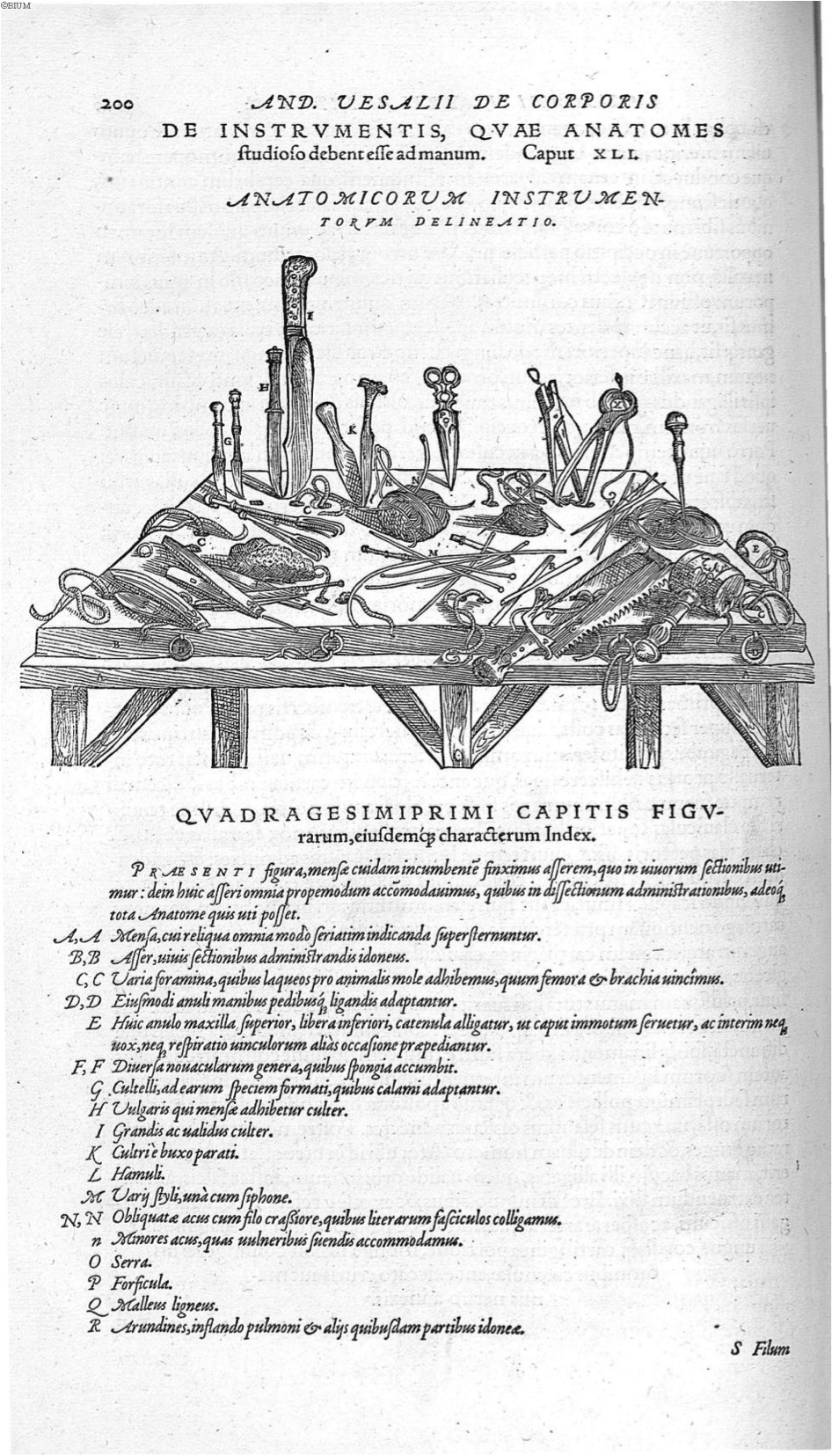
Figure 27. Vesalius, A., de Humani corporis fabrica Libri septem (p. 200), 1543. Link |
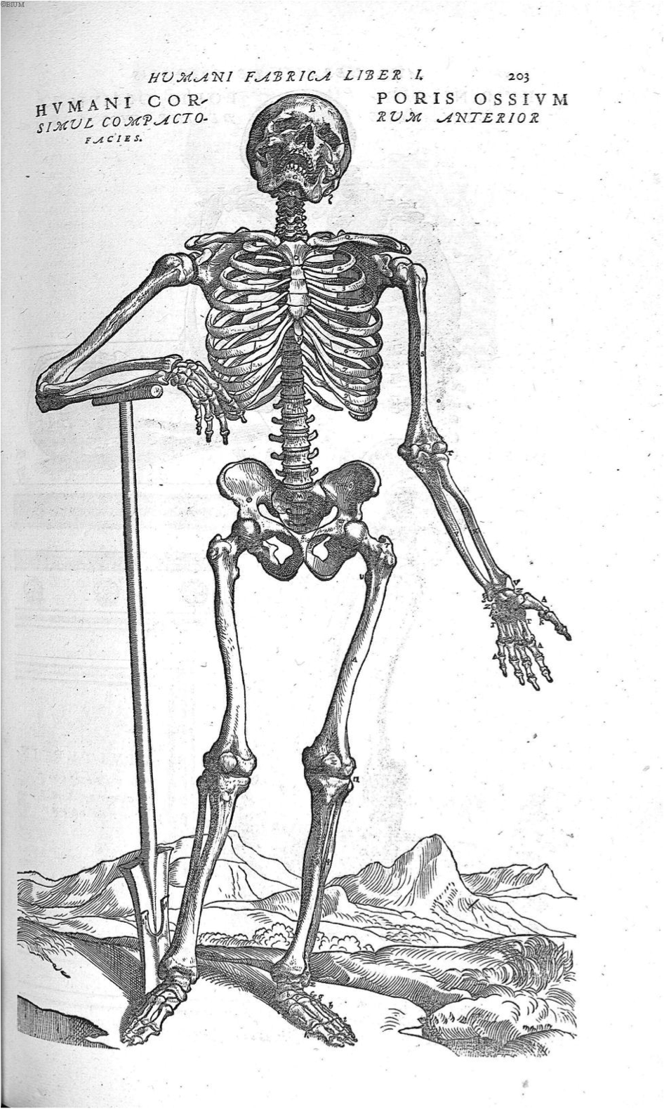
Figure 28. Vesalius, A., de Humani corporis fabrica Libri septem (p. 201), 1543. Link |
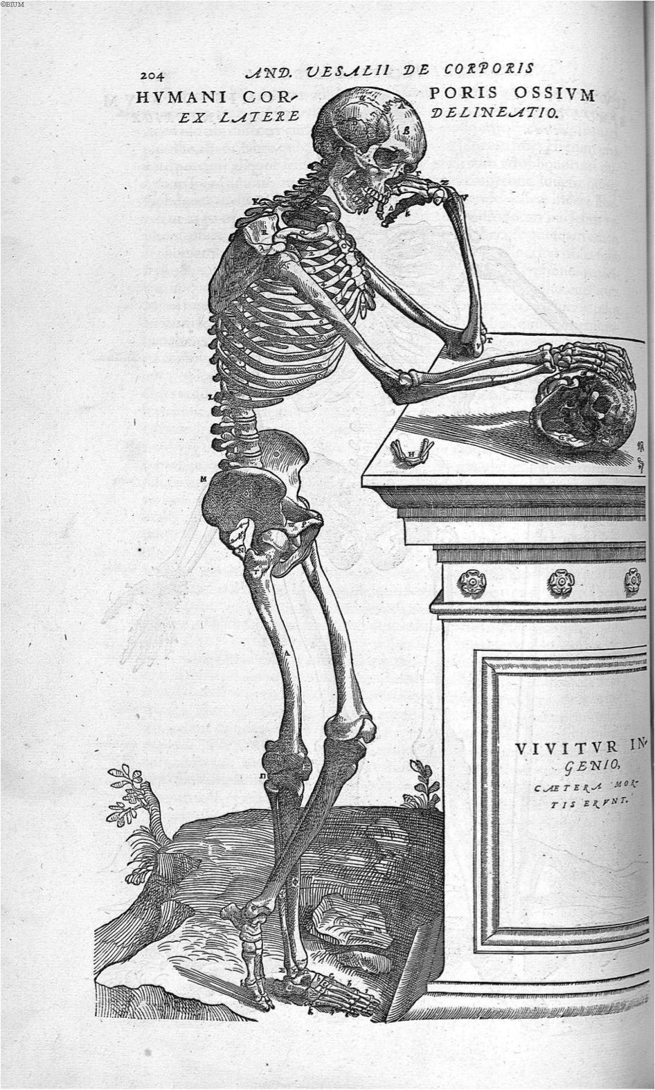
Figure 29. Vesalius, A., de Humani corporis fabrica Libri septem (p. 203), 1543. Link |

Figure 30. Vesalius, A., de Humani corporis fabrica Libri septem (p. 204), 1543. Link |
Book 2: The Ligaments and Muscles
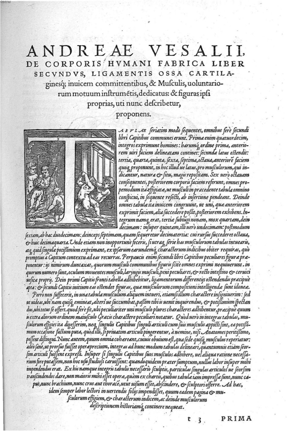
Figure 1. Vesalius, A., de Humani corporis fabrica Libri septem (p. 209), 1543. Link |
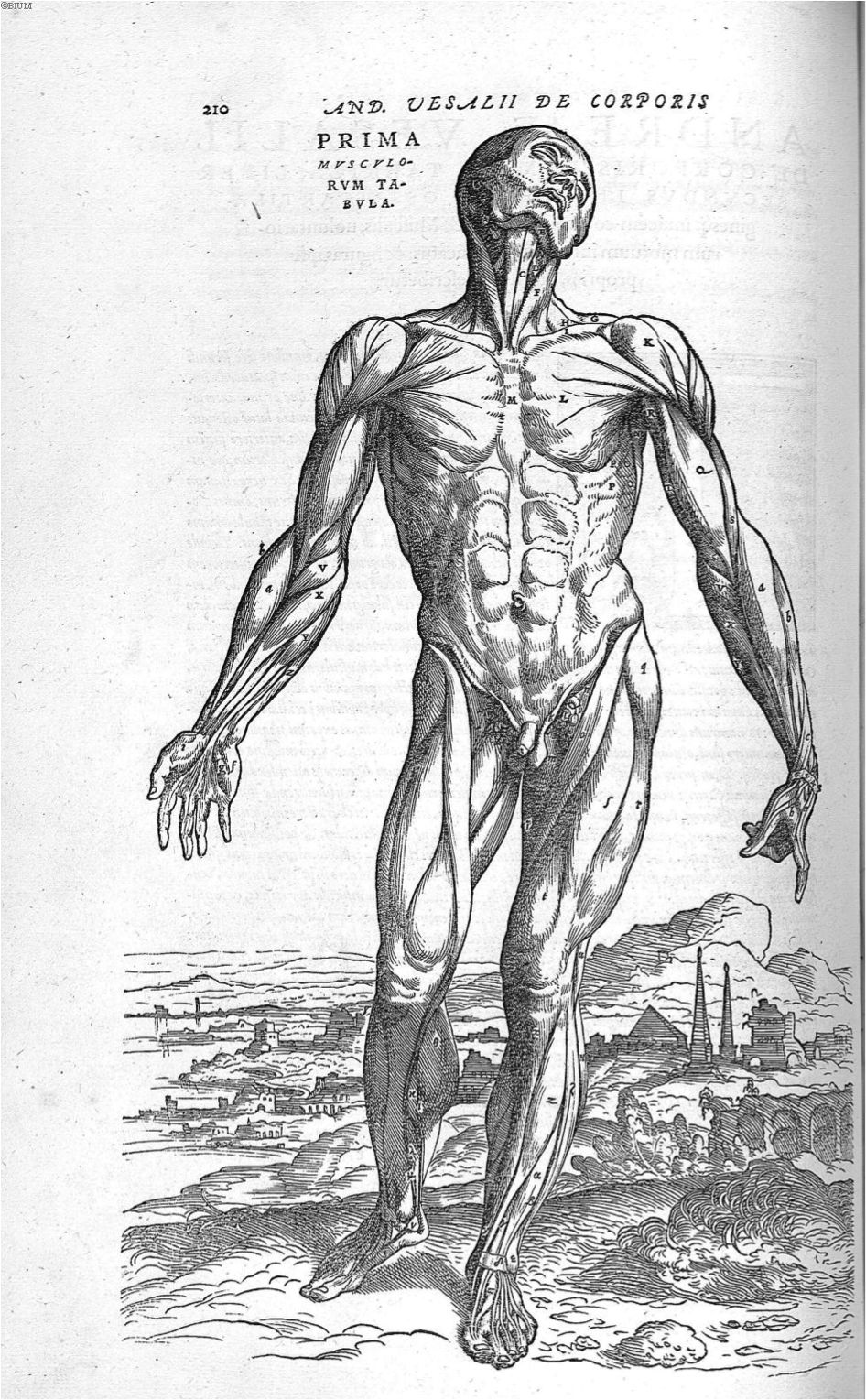
Figure 2. Vesalius, A., de Humani corporis fabrica Libri septem (p. 210), 1543. Link |
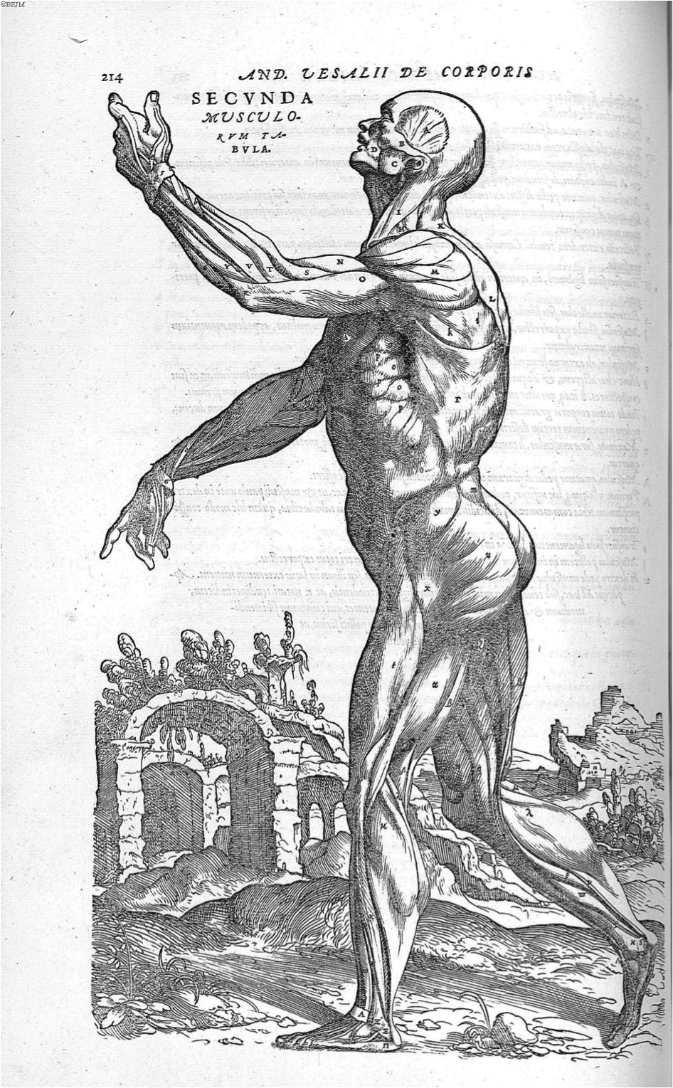
Figure 3. Vesalius, A., de Humani corporis fabrica Libri septem (p. 214), 1543. Link |
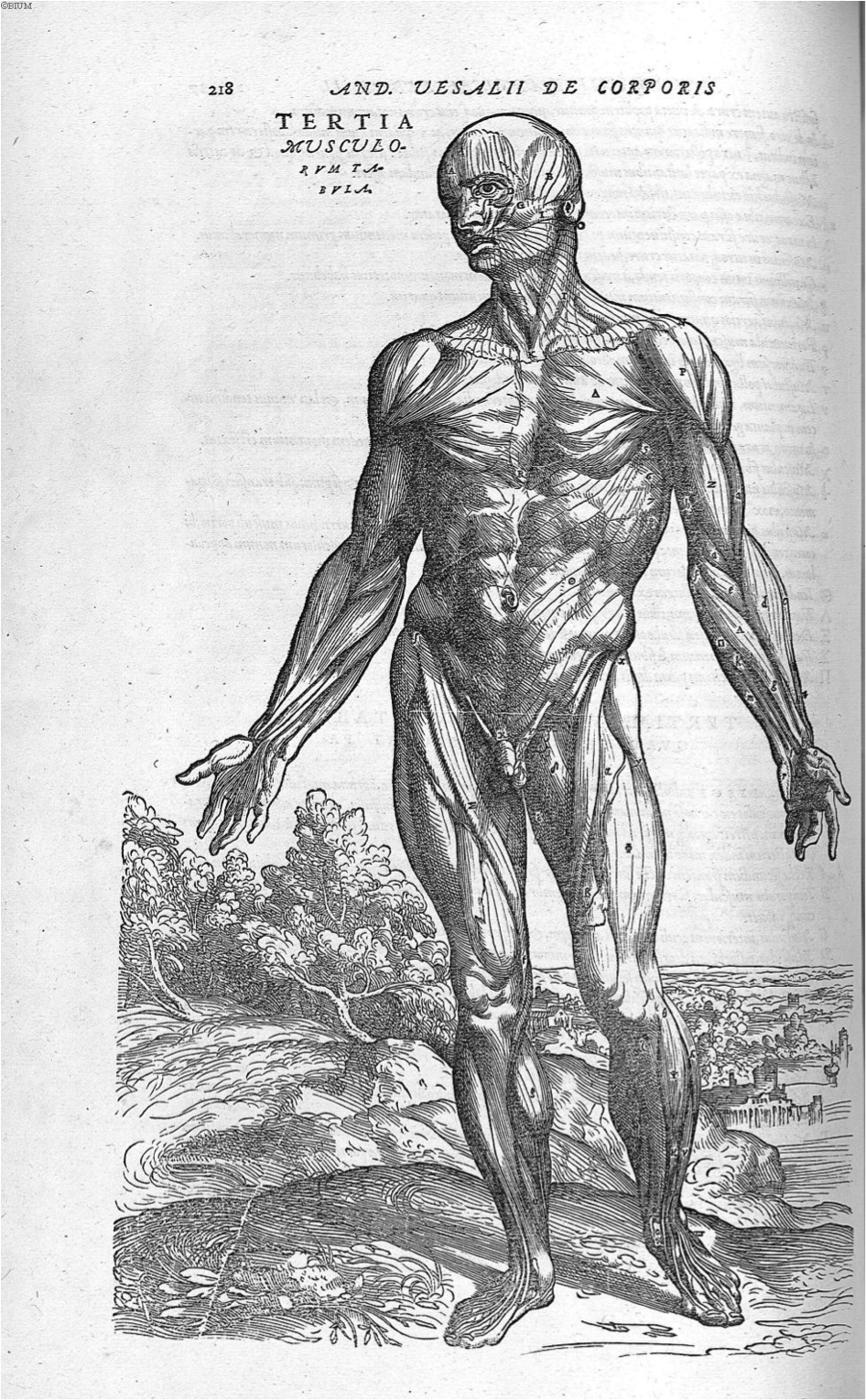
Figure 4. Vesalius, A., de Humani corporis fabrica Libri septem (p. 218), 1543. Link |
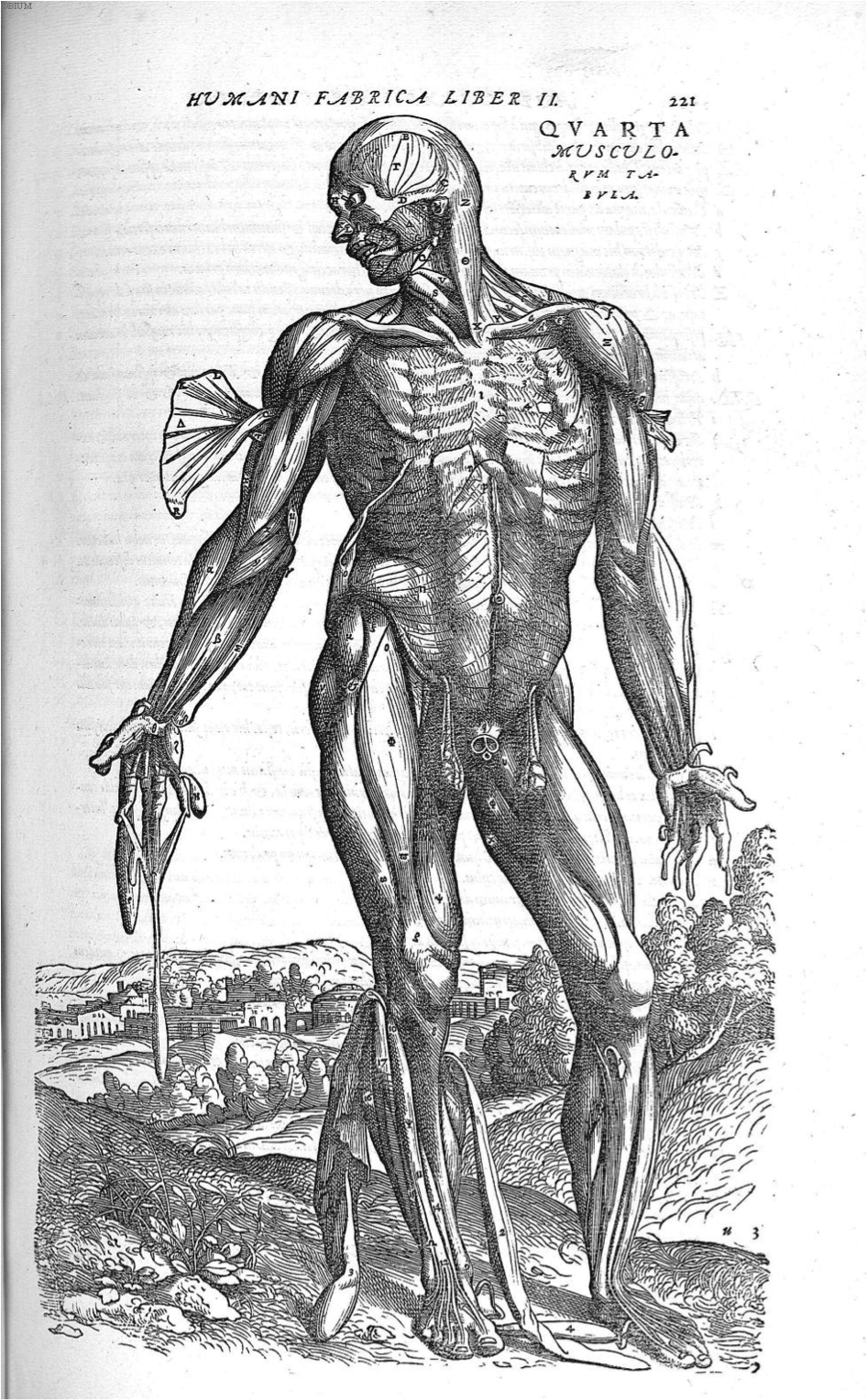
Figure 5. Vesalius, A., de Humani corporis fabrica Libri septem (p. 221), 1543. Link |
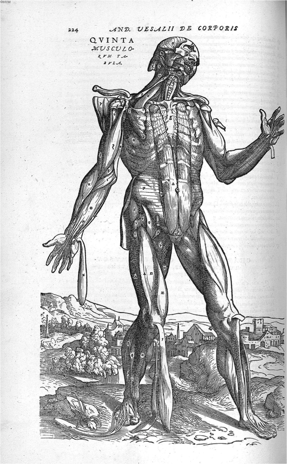
Figure 6. Vesalius, A., de Humani corporis fabrica Libri septem (p. 224), 1543. Link |
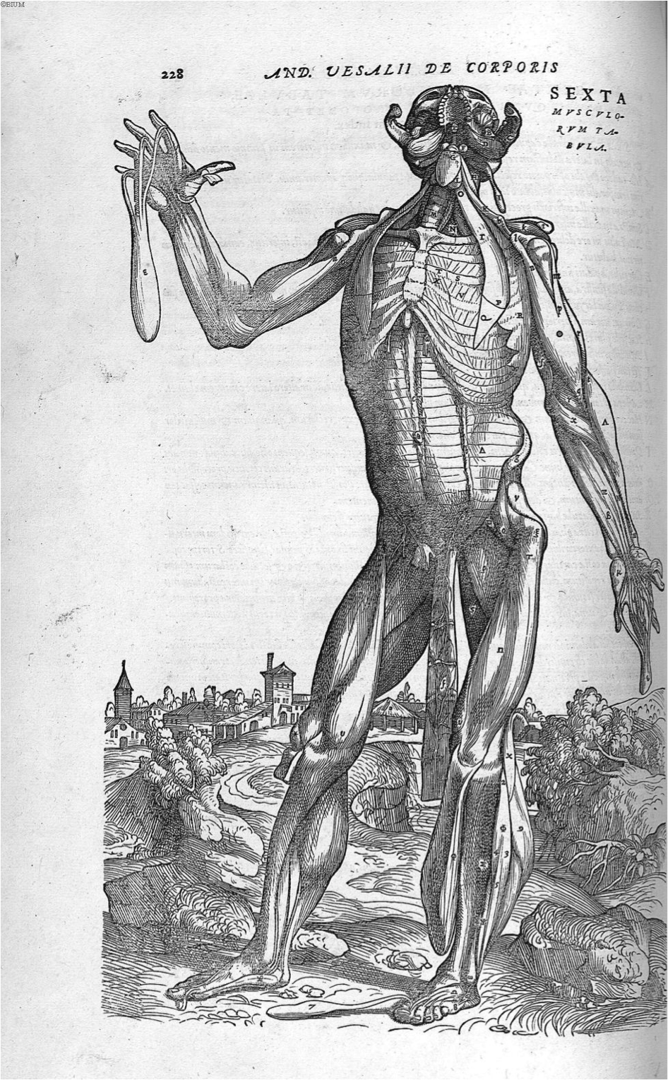
Figure 7. Vesalius, A., de Humani corporis fabrica Libri septem (p. 228), 1543. Link |
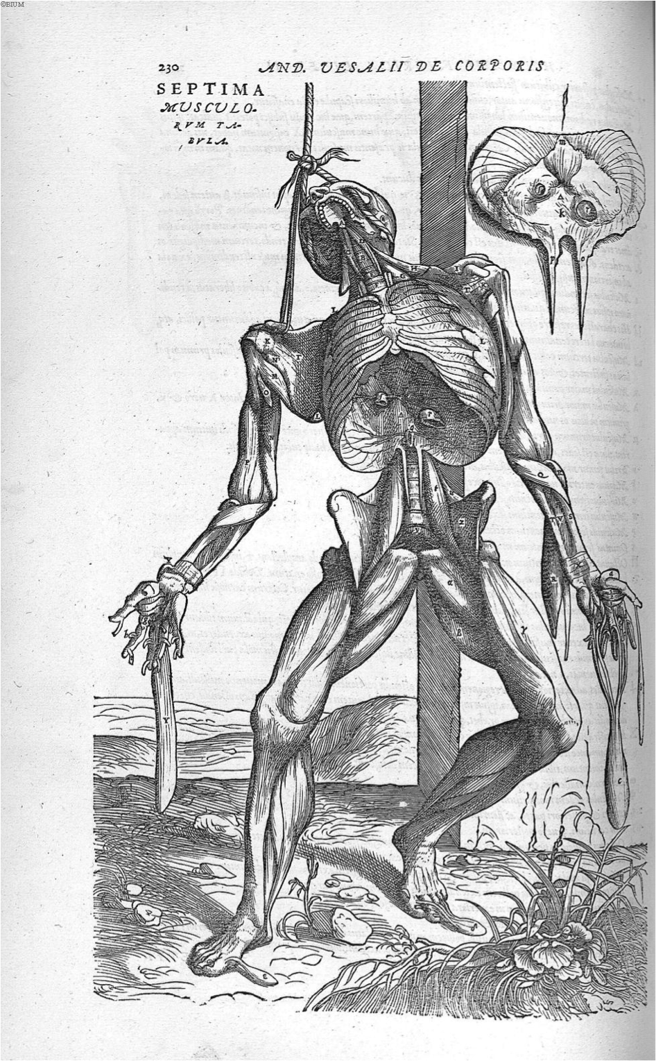
Figure 8. Vesalius, A., de Humani corporis fabrica Libri septem (p. 230), 1543. Link |
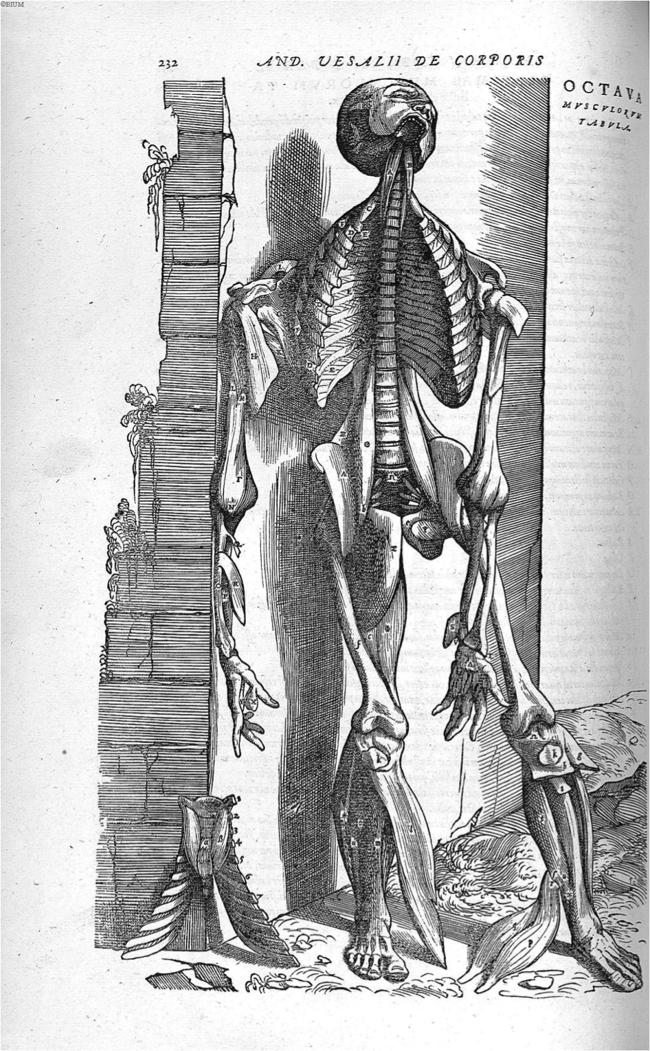
Figure 9. Vesalius, A., de Humani corporis fabrica Libri septem (p. 232), 1543. Link |
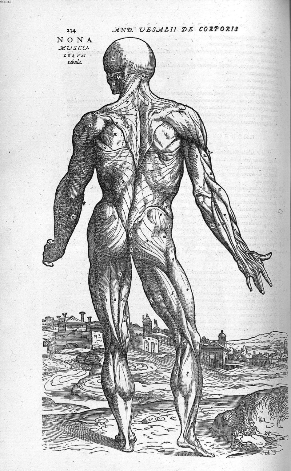
Figure 10. Vesalius, A., de Humani corporis fabrica Libri septem (p. 234), 1543. Link |
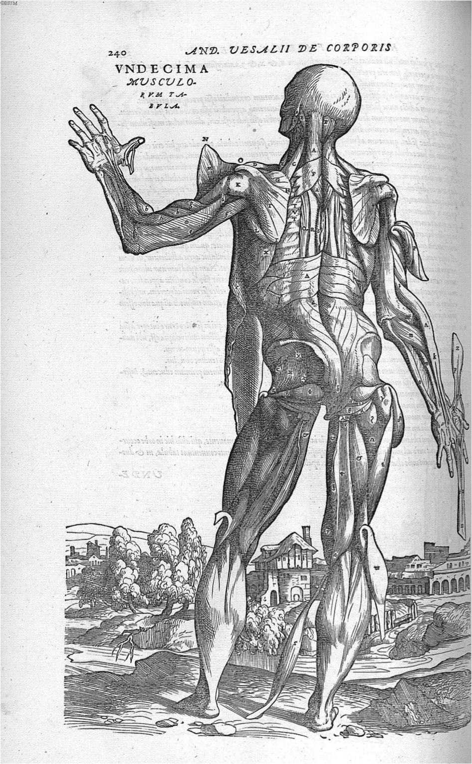
Figure 11. Vesalius, A., de Humani corporis fabrica Libri septem (p. 2408), 1543. Link |
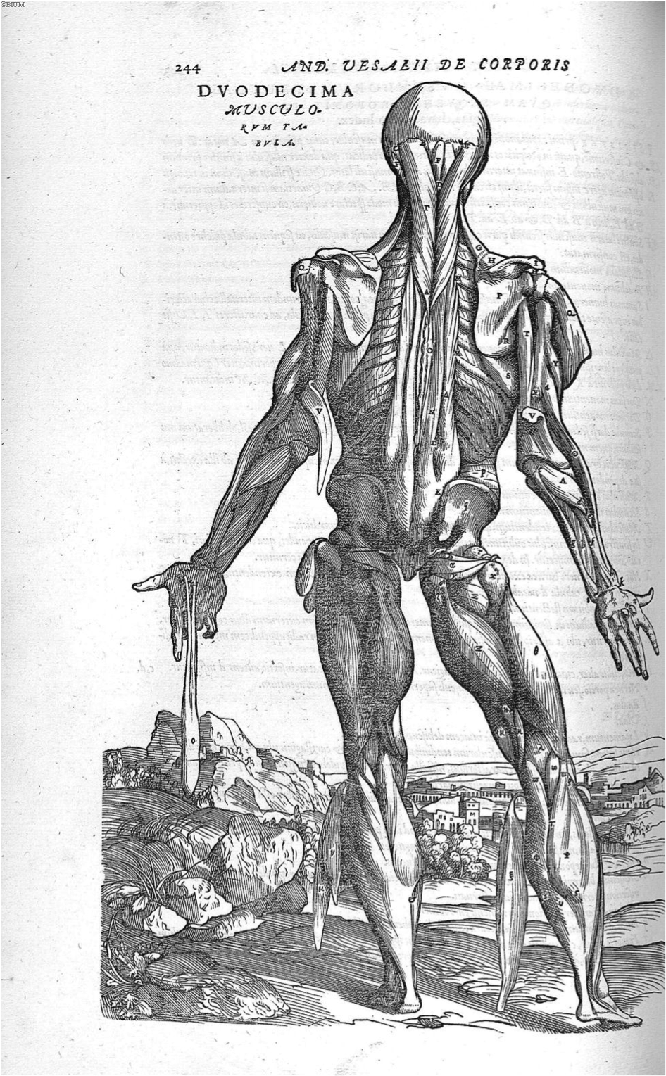
Figure 12. Vesalius, A., de Humani corporis fabrica Libri septem (p. 244), 1543. Link |
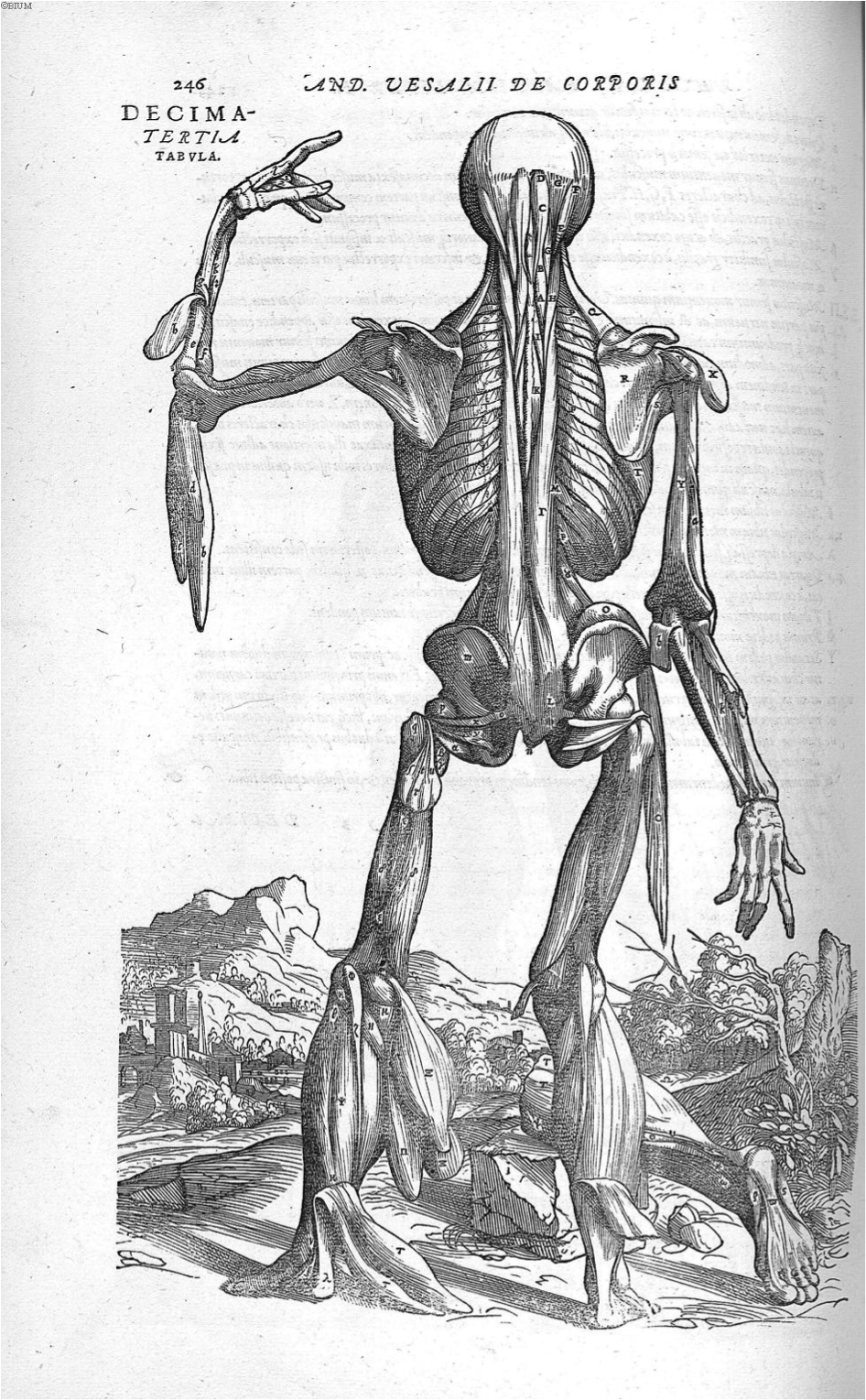
Figure 13. Vesalius, A., de Humani corporis fabrica Libri septem (p. 246), 1543. Link |
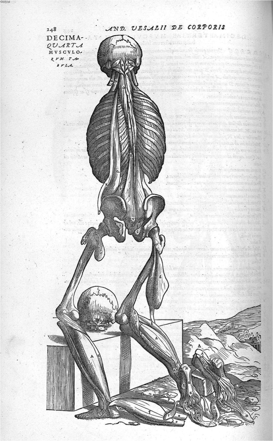
Figure 14. Vesalius, A., de Humani corporis fabrica Libri septem (p. 248), 1543. Link |
Book 3: The Veins and Arteries
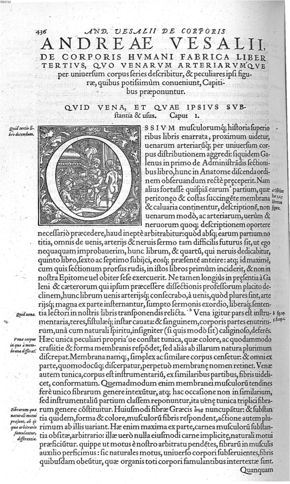
Figure 1. Vesalius, A., de Humani corporis fabrica Libri septem (p. 436), 1543. Link |
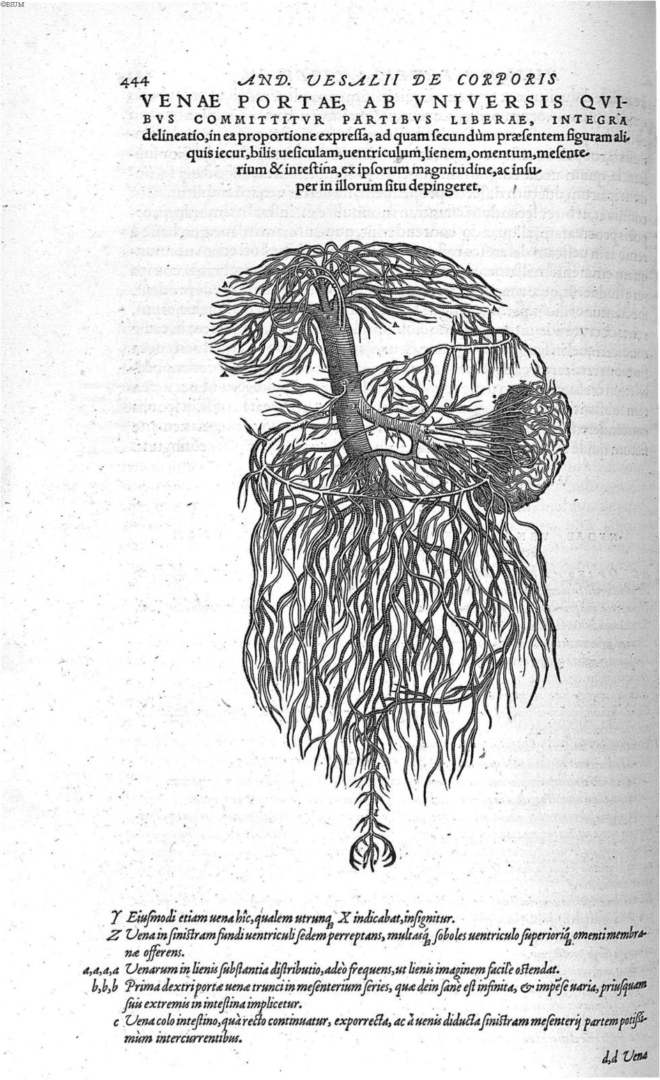
Figure 2. Vesalius, A., de Humani corporis fabrica Libri septem (p. 444), 1543. Link |

Figure 3. Vesalius, A., de Humani corporis fabrica Libri septem (p. 450), 1543. Link |
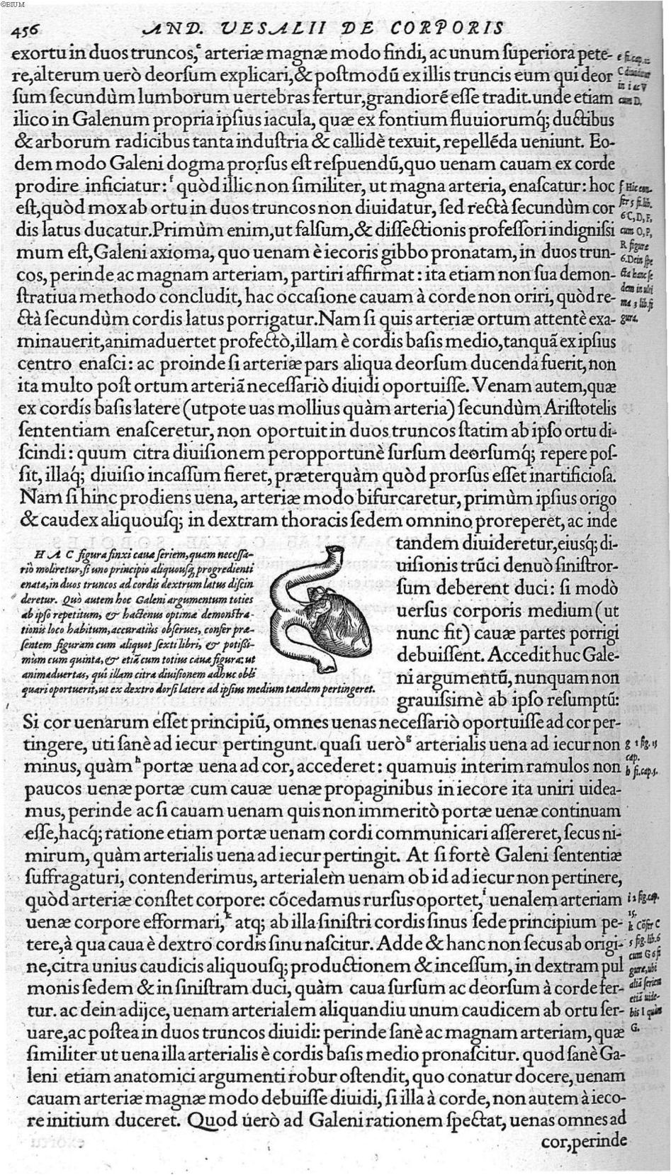
Figure 4. Vesalius, A., de Humani corporis fabrica Libri septem (p. 456), 1543. Link |
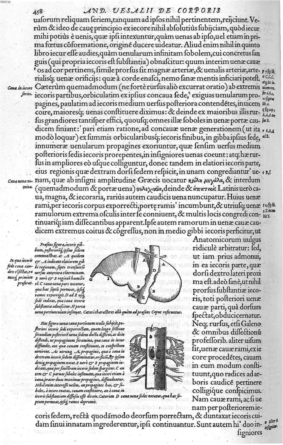
Figure 5. Vesalius, A., de Humani corporis fabrica Libri septem (p. 458), 1543. Link |
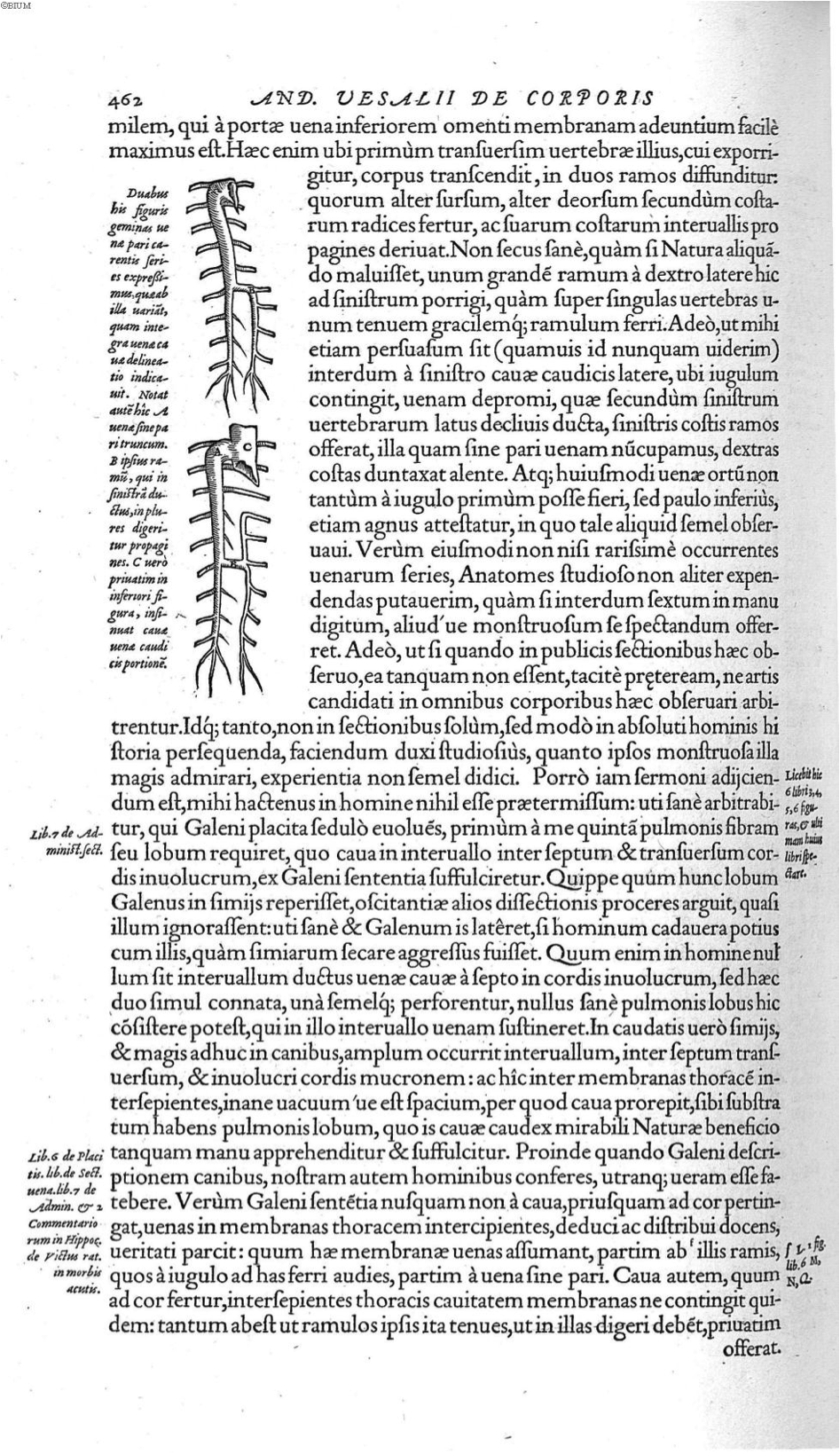
Figure 6. Vesalius, A., de Humani corporis fabrica Libri septem (p. 462), 1543. Link |
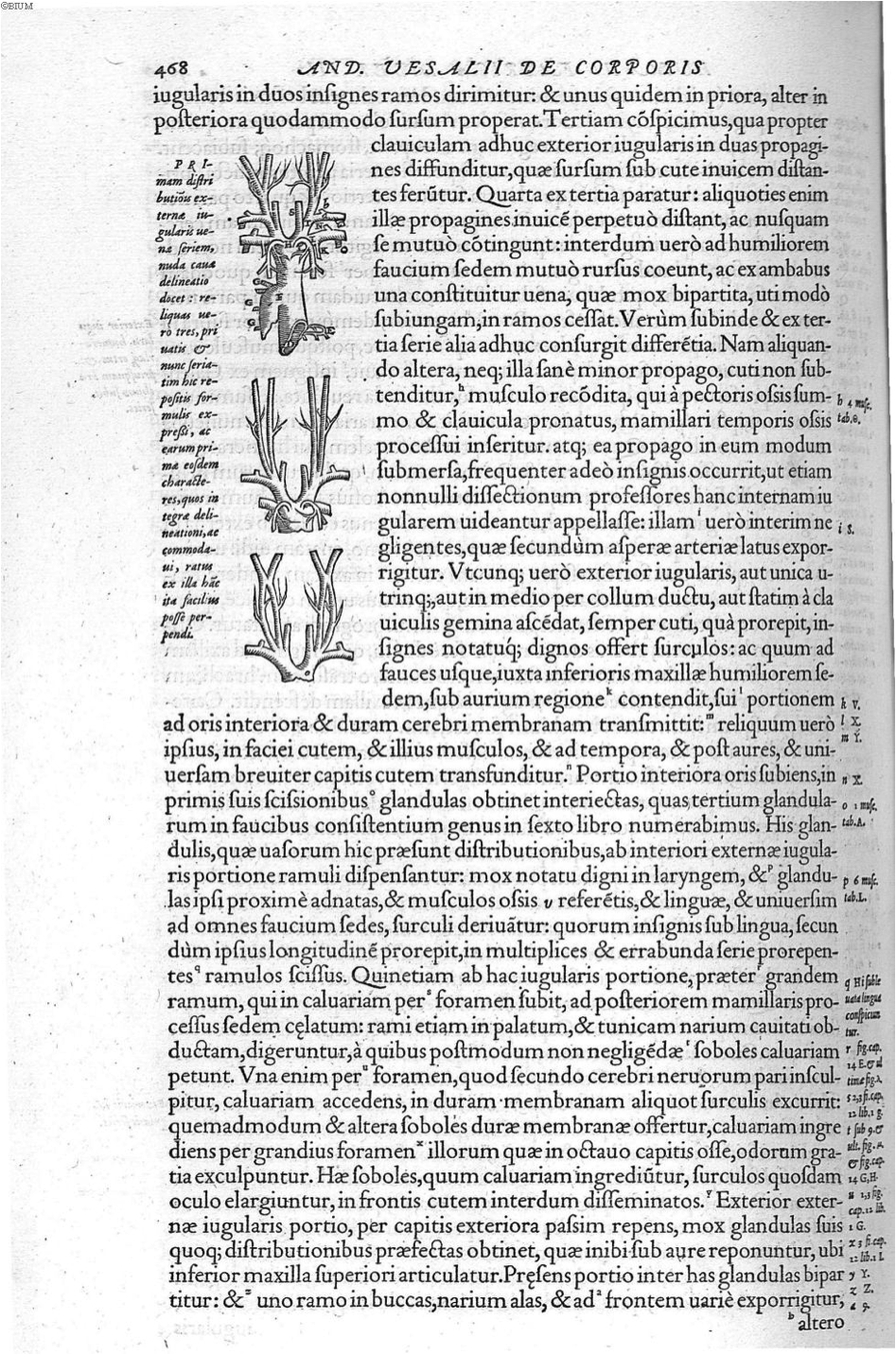
Figure 7. Vesalius, A., de Humani corporis fabrica Libri septem (p. 468), 1543. Link |
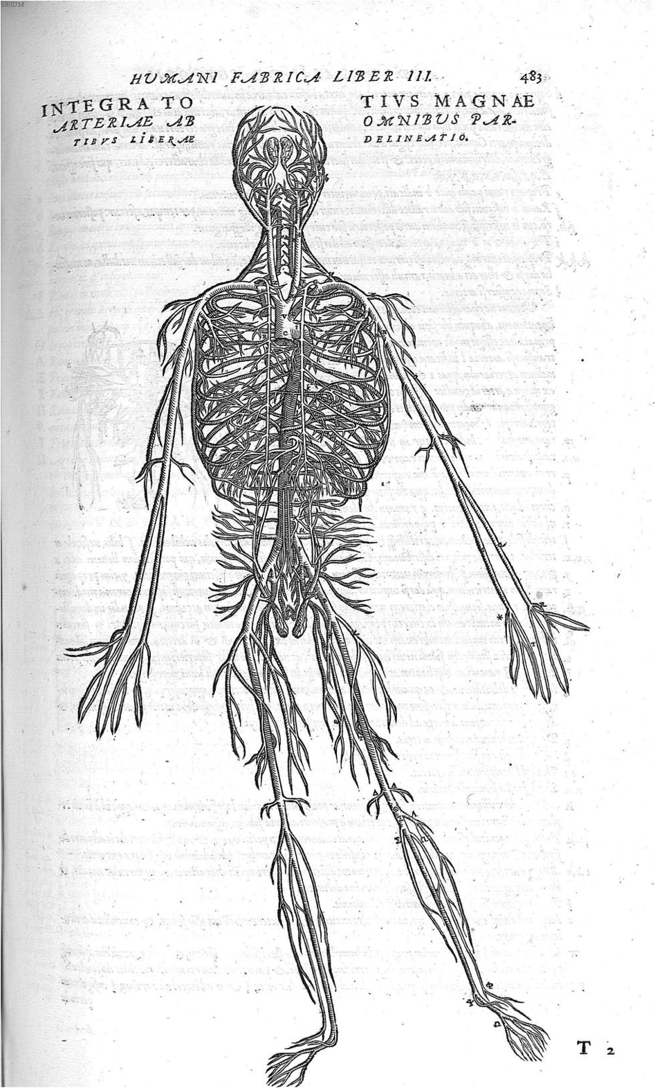
Figure 8. Vesalius, A., de Humani corporis fabrica Libri septem (p. 483), 1543. Link |
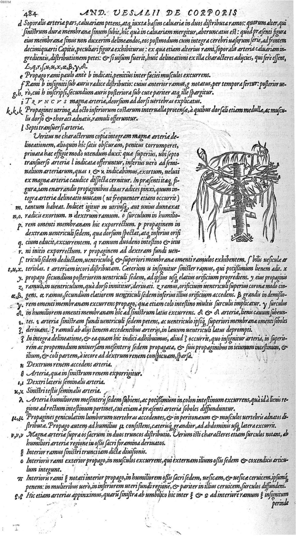
Figure 9. Vesalius, A., de Humani corporis fabrica Libri septem (p. 484), 1543. Link |
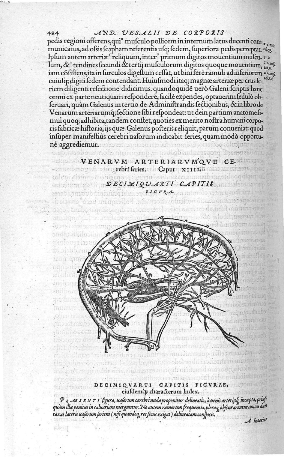
Figure 10. Vesalius, A., de Humani corporis fabrica Libri septem (p. 494), 1543. Link |
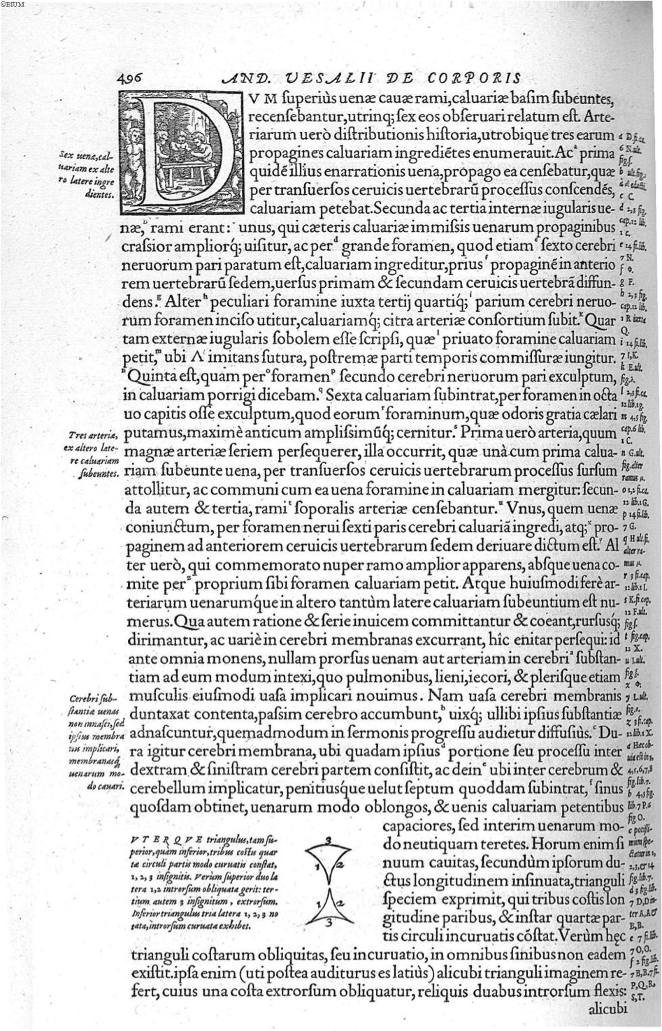
Figure 11. Vesalius, A., de Humani corporis fabrica Libri septem (p. 496), 1543. Link |
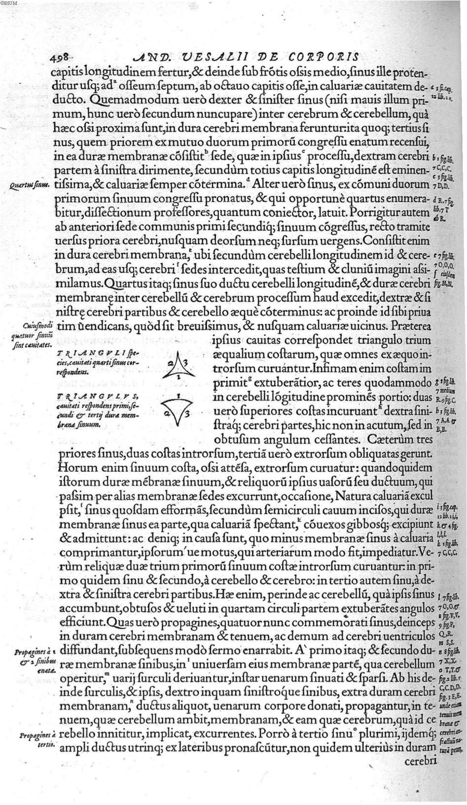
Figure 12. Vesalius, A., de Humani corporis fabrica Libri septem (p. 498), 1543. Link |
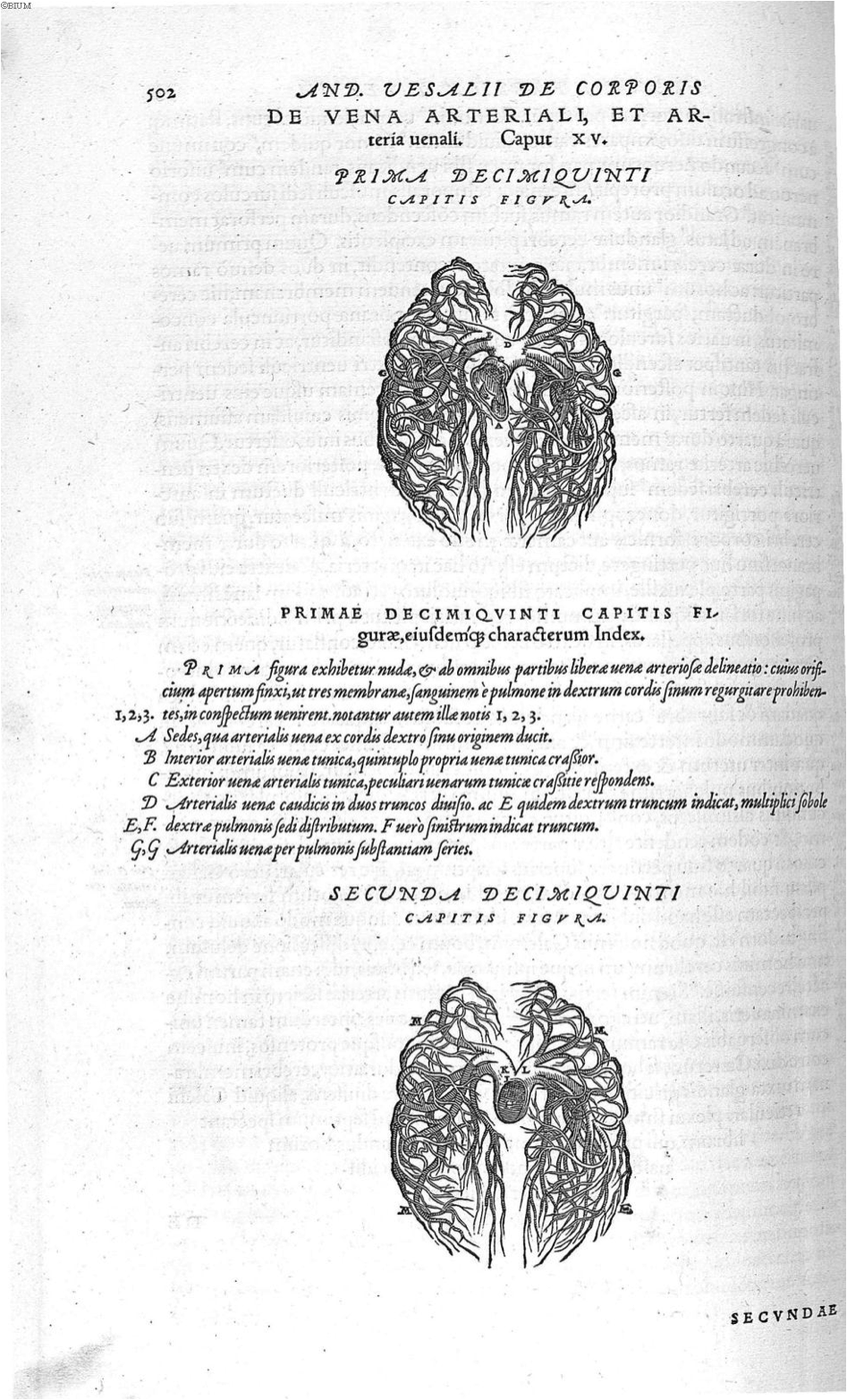
Figure 13. Vesalius, A., de Humani corporis fabrica Libri septem (p. 502), 1543. Link |
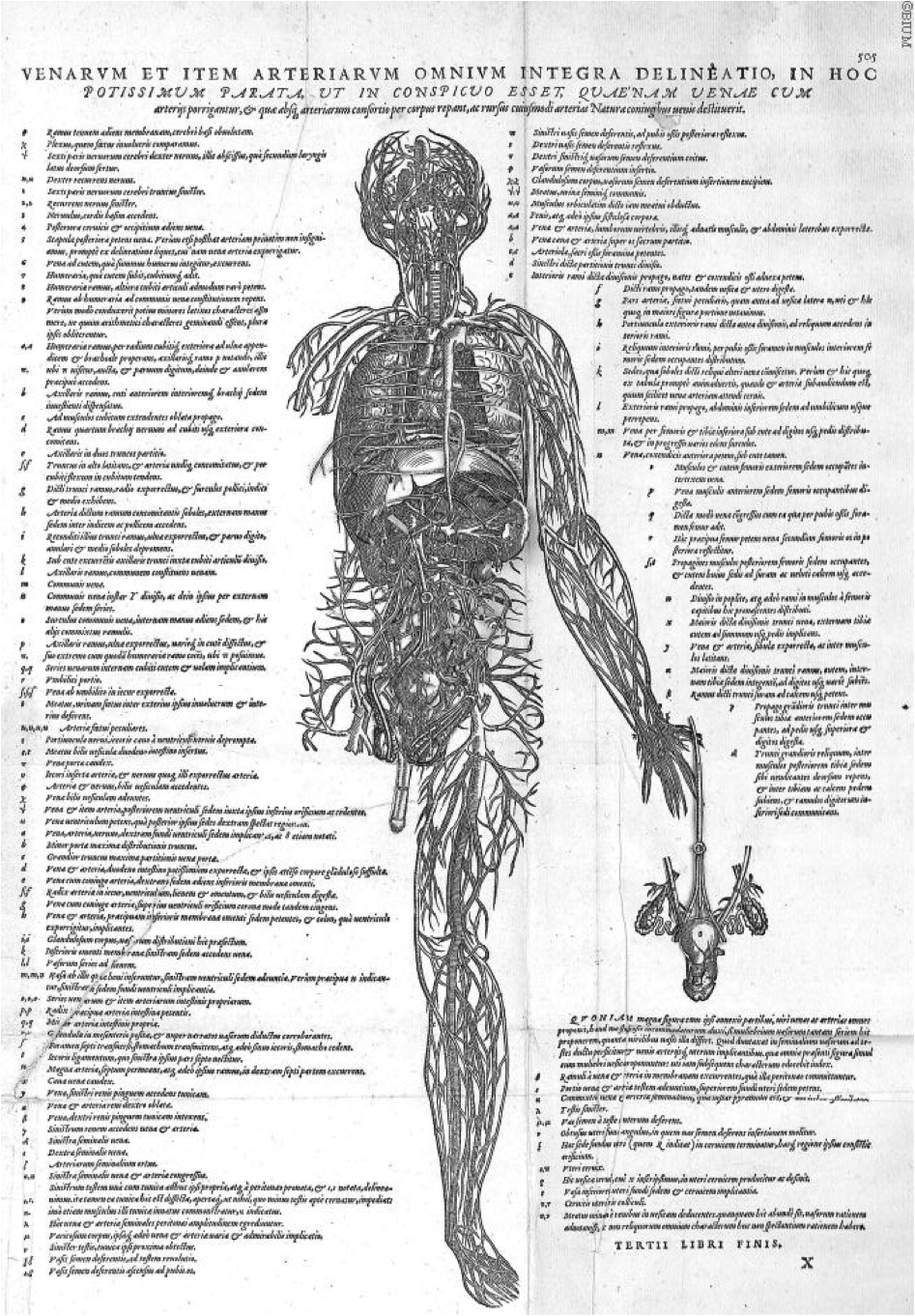
Figure 14. Vesalius, A., de Humani corporis fabrica Libri septem (p. 503), 1543. Link |
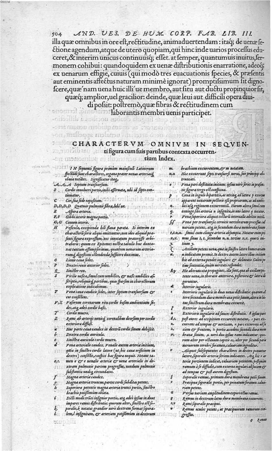
Figure 15. Vesalius, A., de Humani corporis fabrica Libri septem (p. 504), 1543. Link |
Figure 1. Vesalius, A., de Humani corporis fabrica Libri septem (p. 507), 1543. Link |
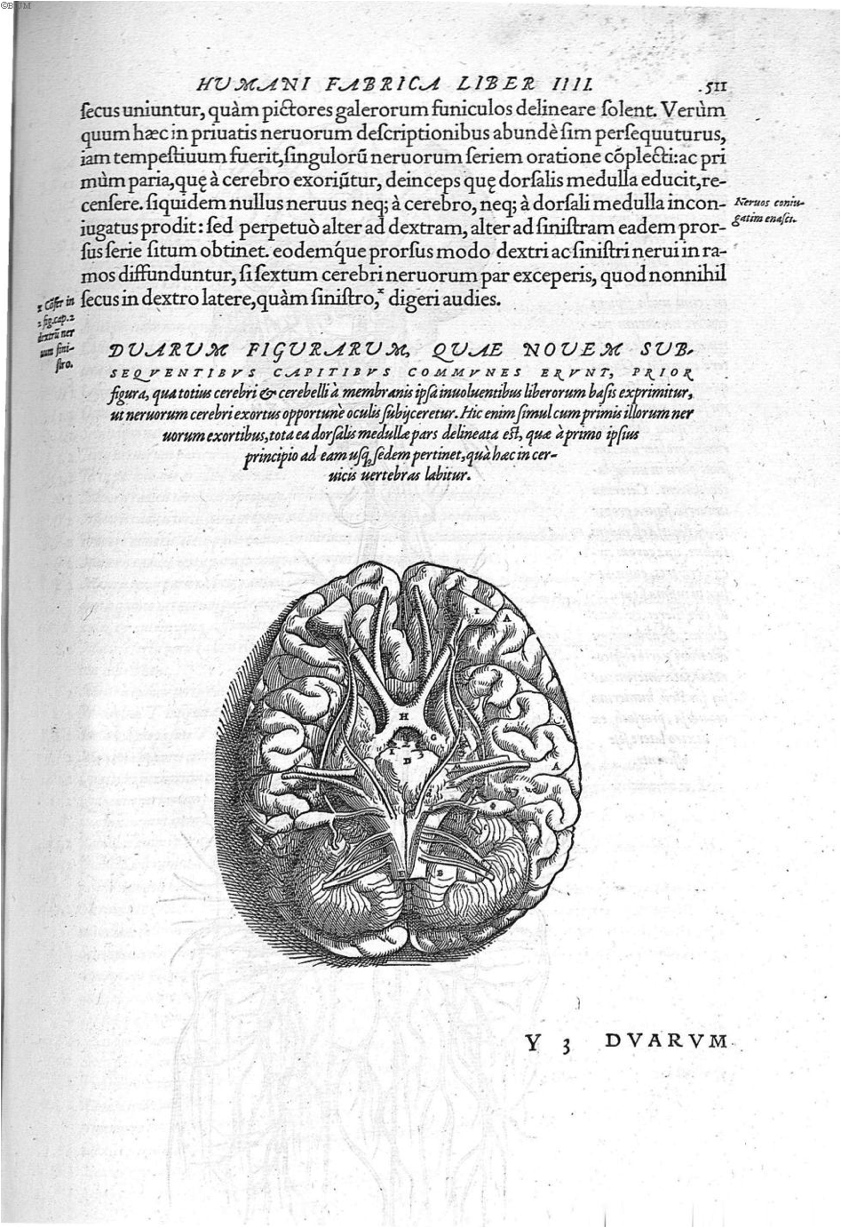
Figure 2. Vesalius, A., de Humani corporis fabrica Libri septem (p. 511), 1543. Link |
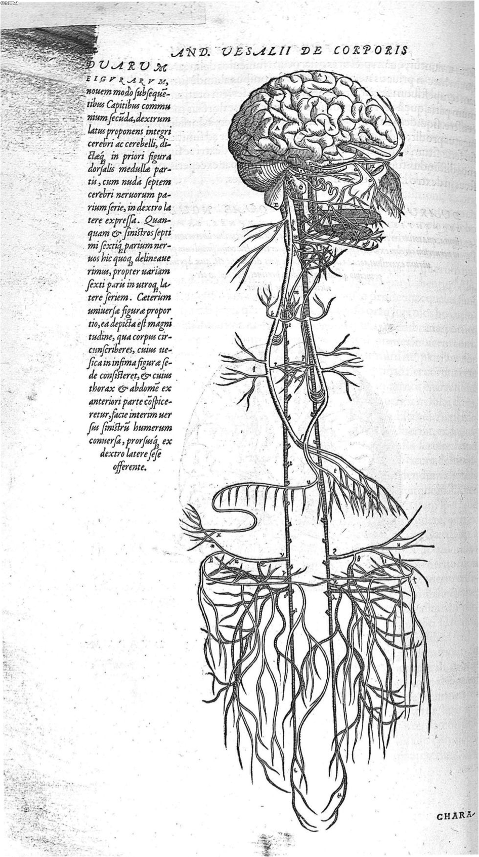
Figure 3. Vesalius, A., de Humani corporis fabrica Libri septem (p. 512), 1543. Link |
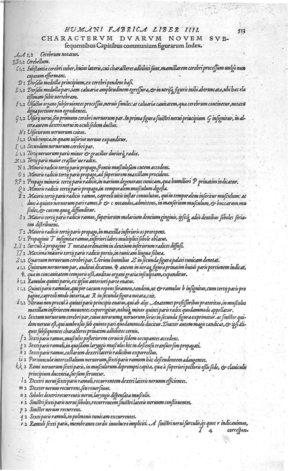
Figure 4. Vesalius, A., de Humani corporis fabrica Libri septem (p. 513), 1543. Link |
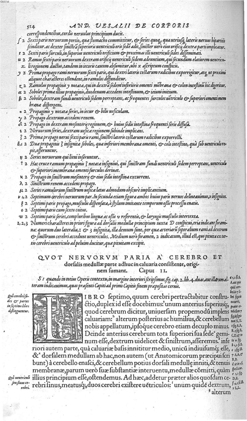
Figure 5. Vesalius, A., de Humani corporis fabrica Libri septem (p. 514), 1543. Link |
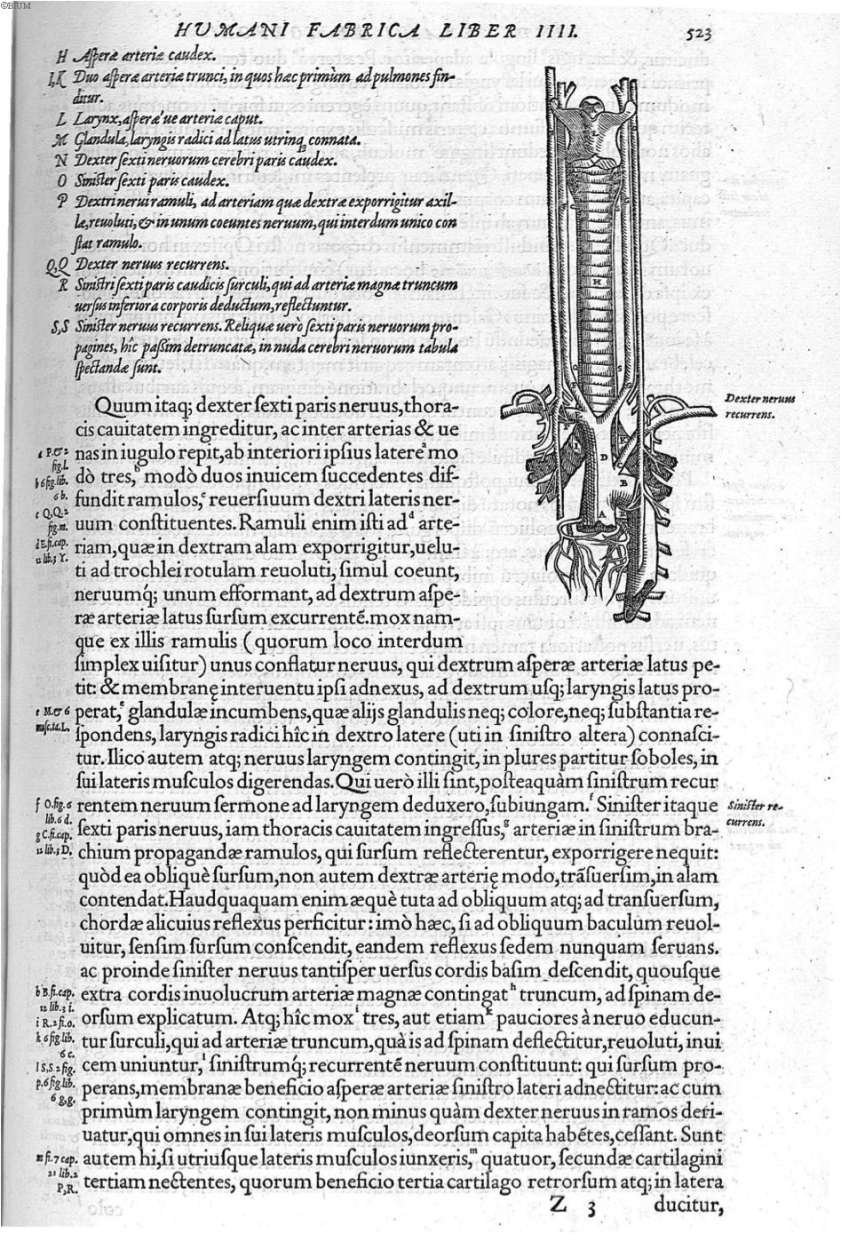
Figure 6. Vesalius, A., de Humani corporis fabrica Libri septem (p. 523), 1543. Link |

Figure 7. Vesalius, A., de Humani corporis fabrica Libri septem (p. 526), 1543. Link |
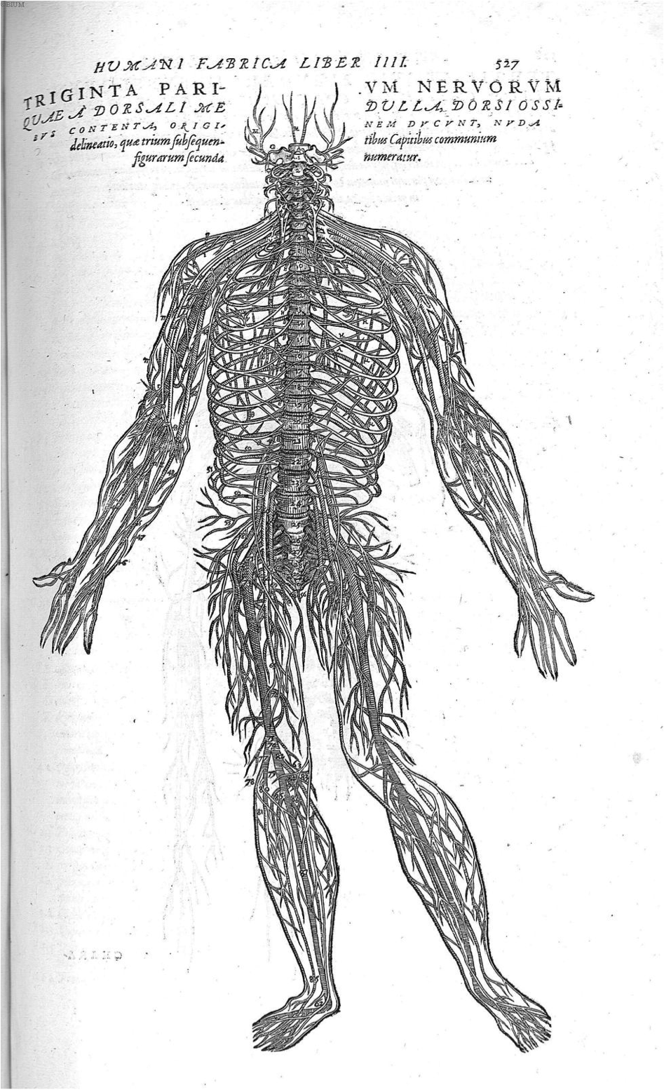
Figure 8. Vesalius, A., de Humani corporis fabrica Libri septem (p. 527), 1543. Link |
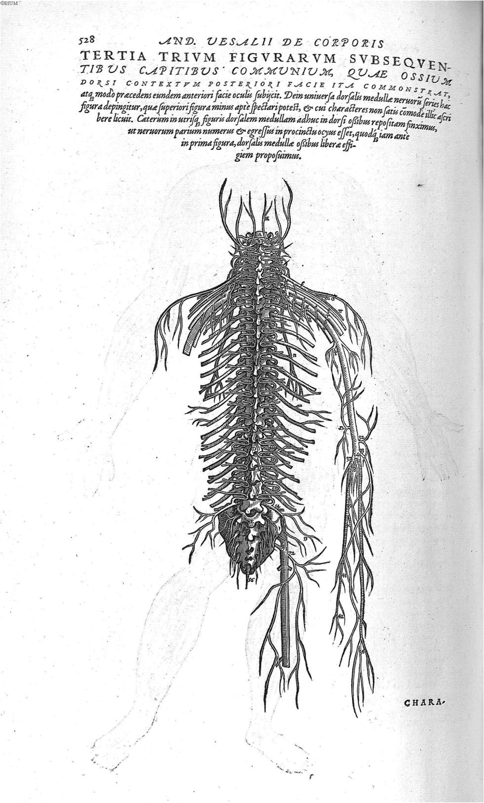
Figure 9. Vesalius, A., de Humani corporis fabrica Libri septem (p. 528), 1543. Link |
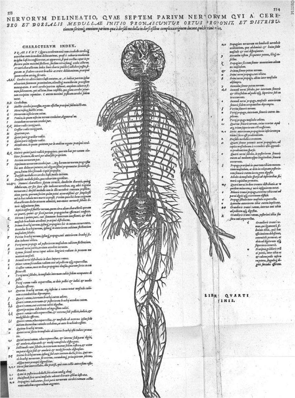
Figure 10. Vesalius, A., de Humani corporis fabrica Libri septem (p. 553-554), 1543. Link |
Book 5: The Organs of Nutrition and Generation
Book 6: The Heart and Associated Organs
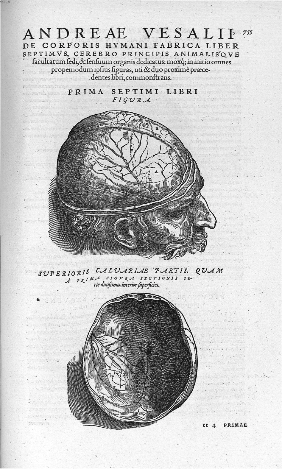
Figure 1. Vesalius, A., de Humani corporis fabrica Libri septem (p. 755), 1543. Link |
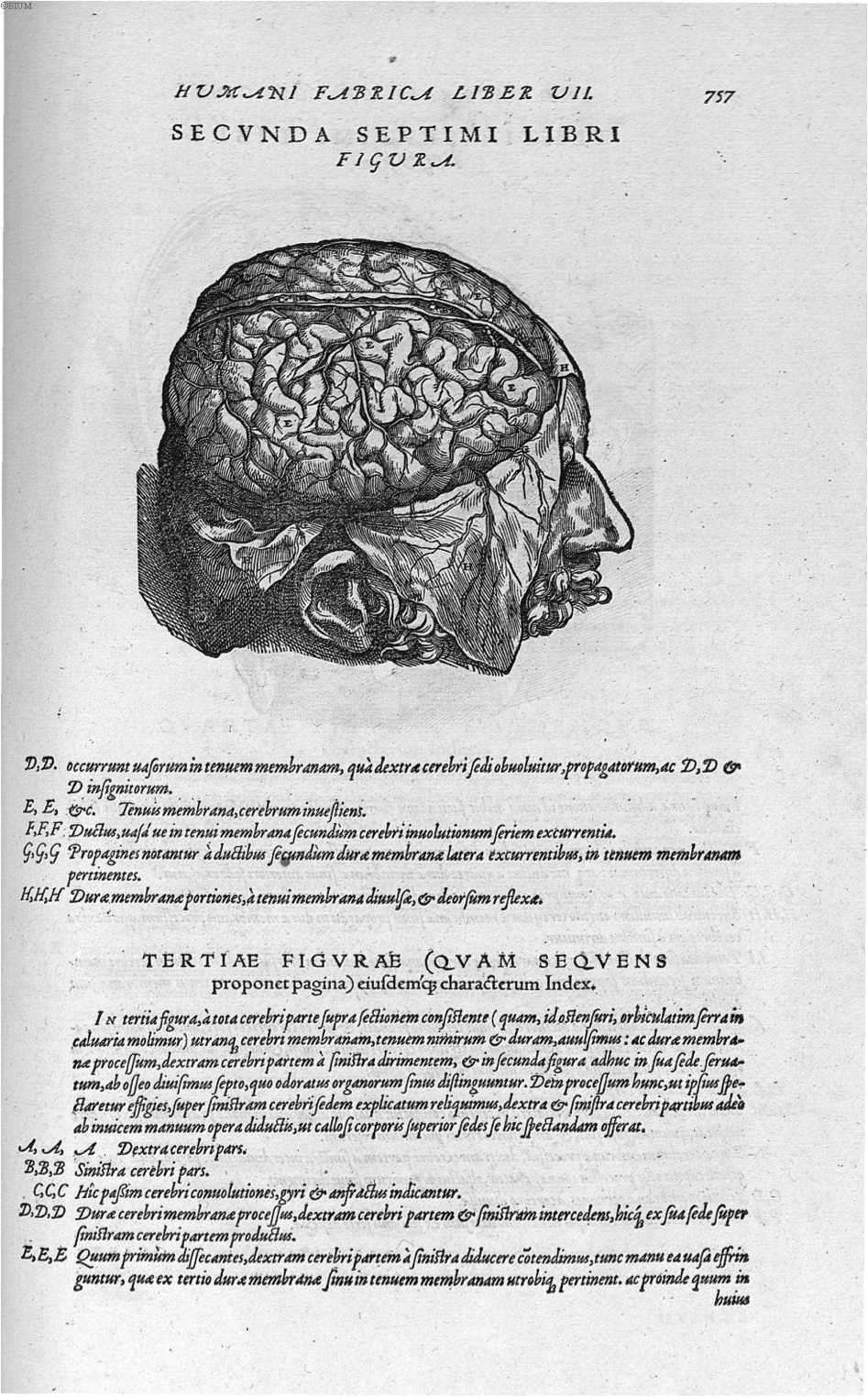
Figure 2. Vesalius, A., de Humani corporis fabrica Libri septem (p. 757), 1543. Link |
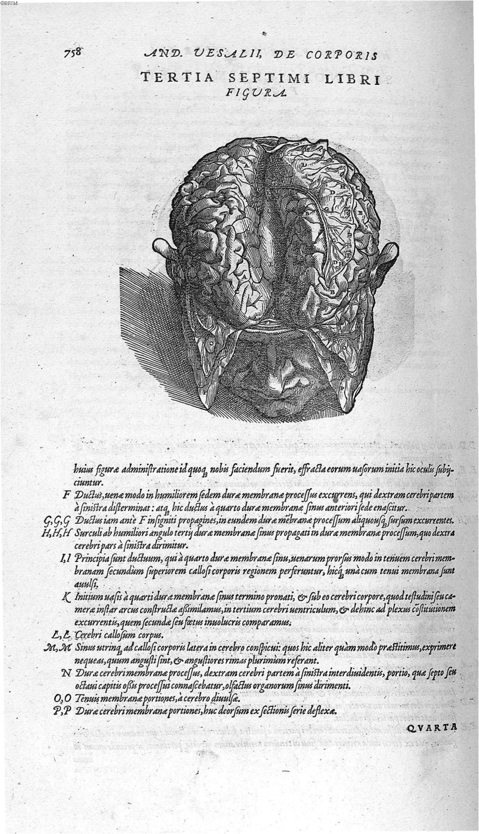
Figure 3. Vesalius, A., de Humani corporis fabrica Libri septem (p. 758), 1543. Link |
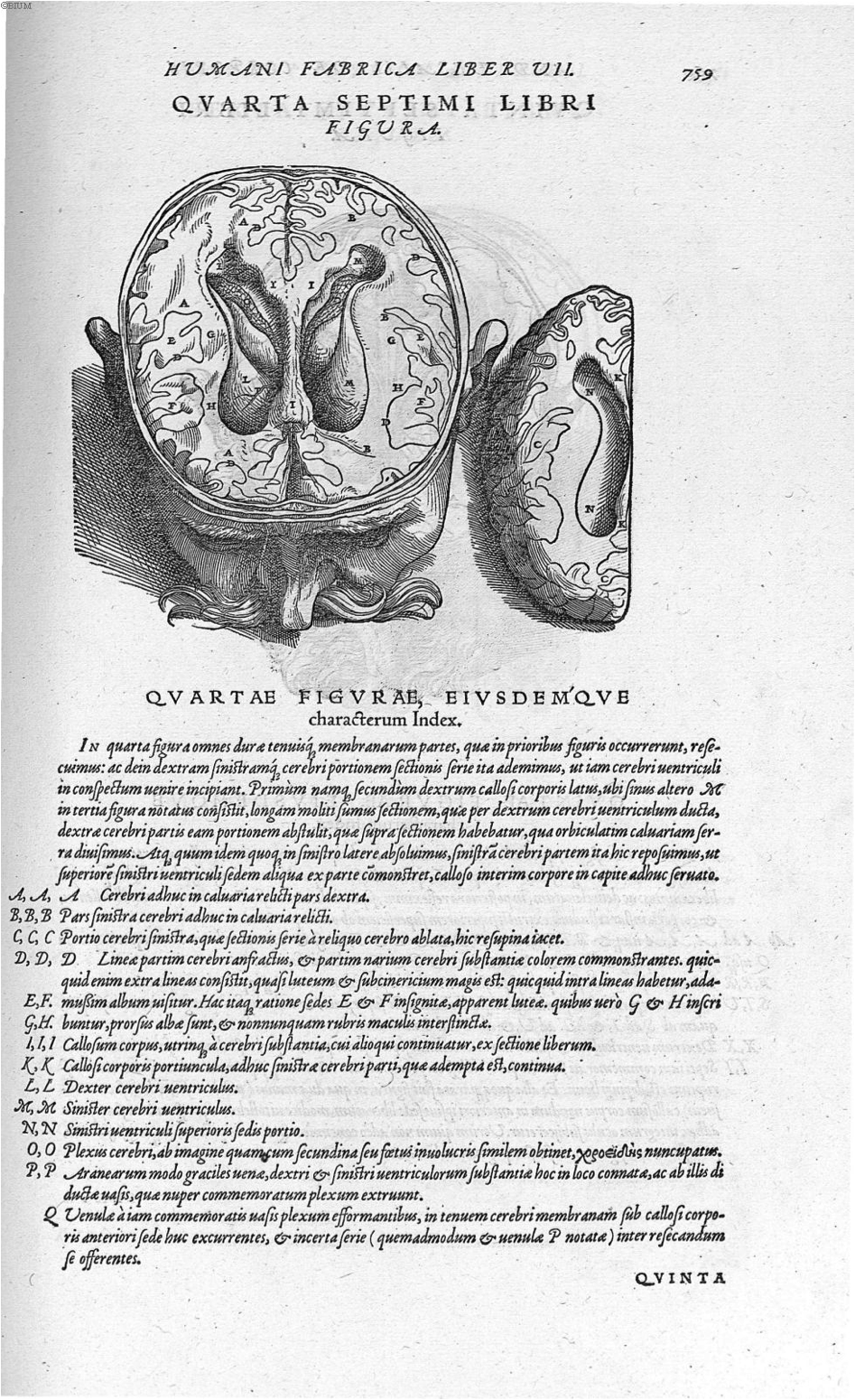
Figure 4. Vesalius, A., de Humani corporis fabrica Libri septem (p. 759), 1543. Link |
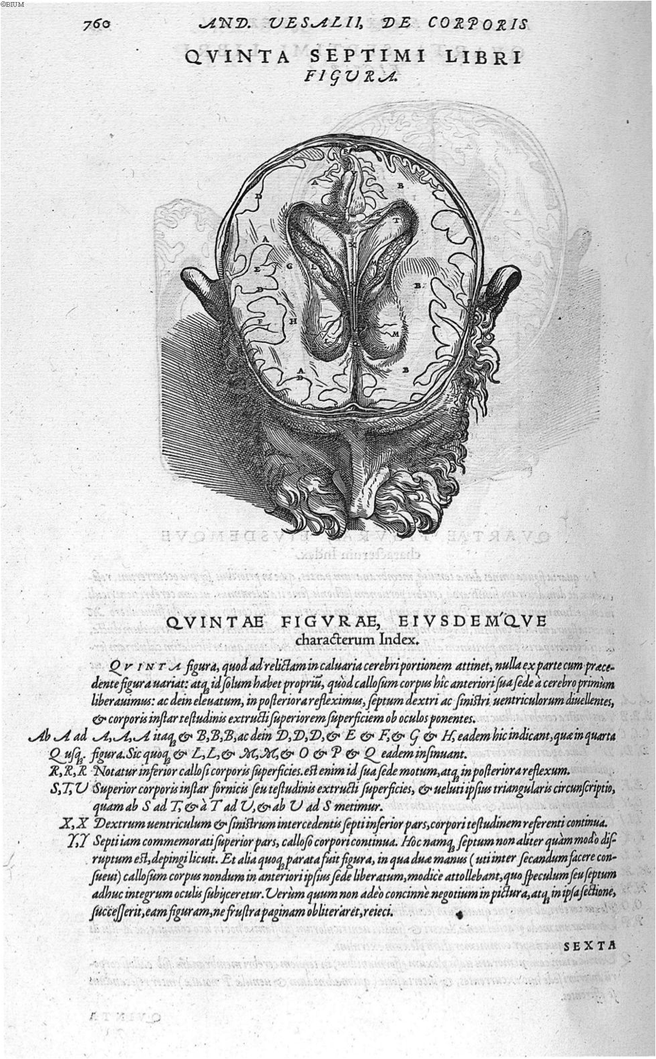
Figure 5. Vesalius, A., de Humani corporis fabrica Libri septem (p. 760), 1543. Link |
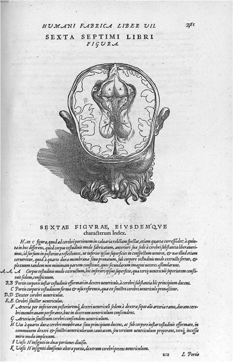
Figure 6. Vesalius, A., de Humani corporis fabrica Libri septem (p. 761), 1543. Link |
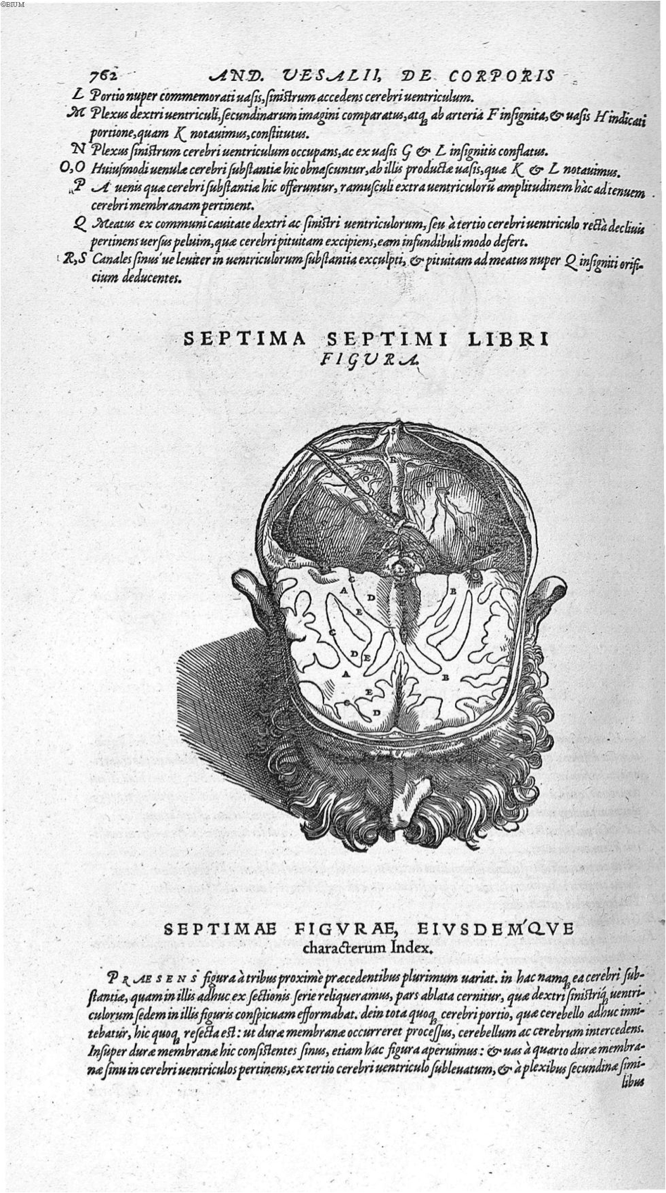
Figure 7. Vesalius, A., de Humani corporis fabrica Libri septem (p. 762), 1543. Link |
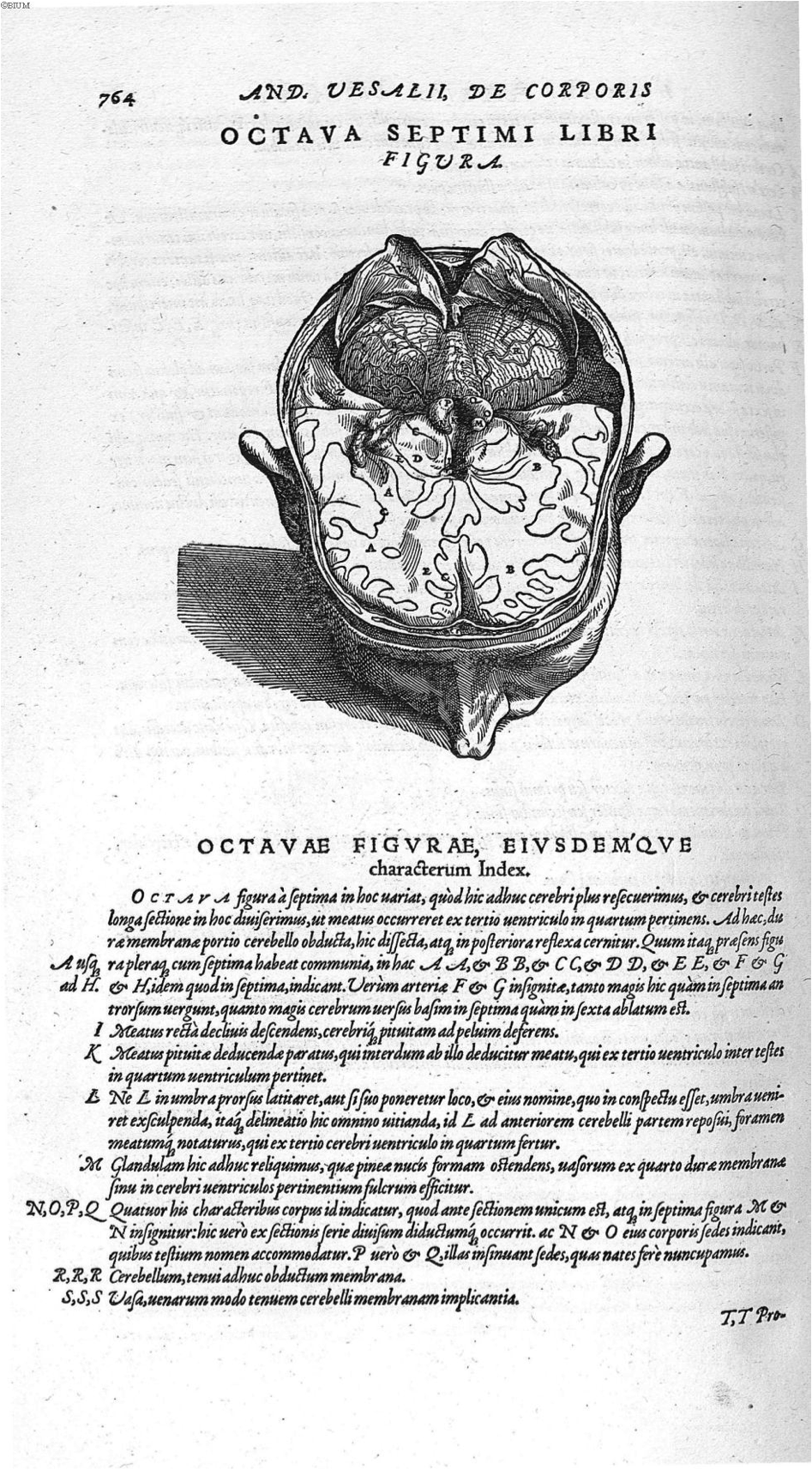
Figure 8. Vesalius, A., de Humani corporis fabrica Libri septem (p. 764), 1543. Link |
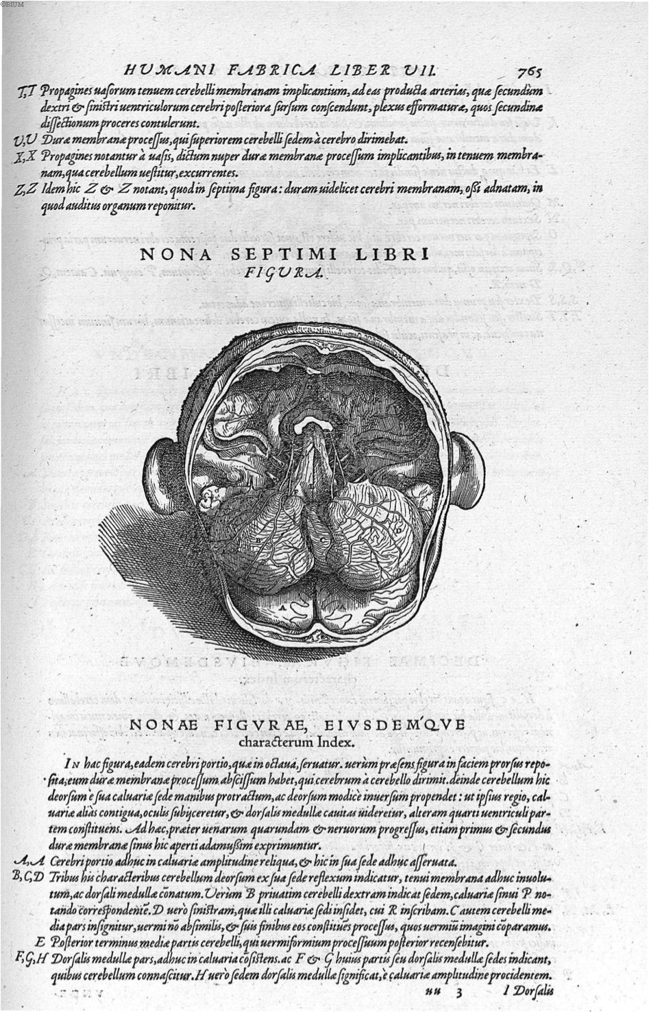
Figure 9. Vesalius, A., de Humani corporis fabrica Libri septem (p. 765), 1543. Link |
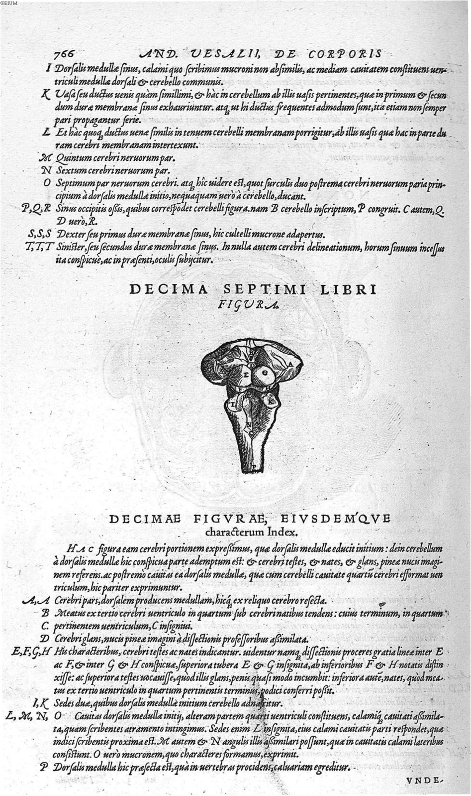
Figure 10. Vesalius, A., de Humani corporis fabrica Libri septem (p. 766), 1543. Link |
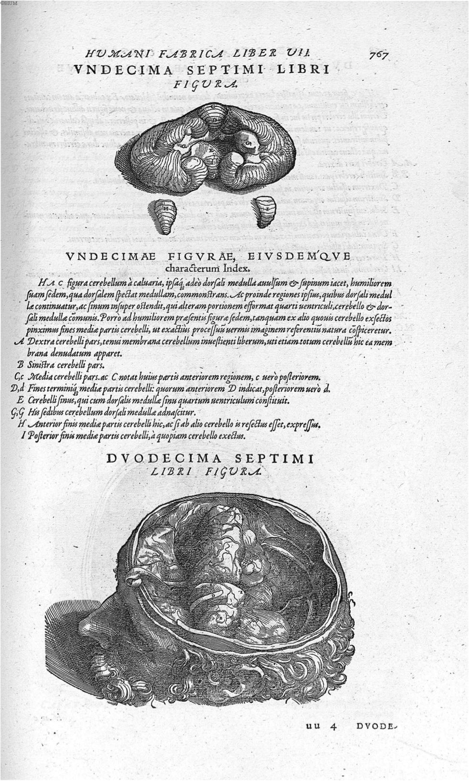
Figure 11. Vesalius, A., de Humani corporis fabrica Libri septem (p. 767), 1543. Link |
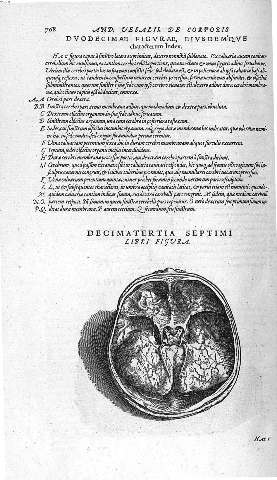
Figure 12. Vesalius, A., de Humani corporis fabrica Libri septem (p. 768), 1543. Link |
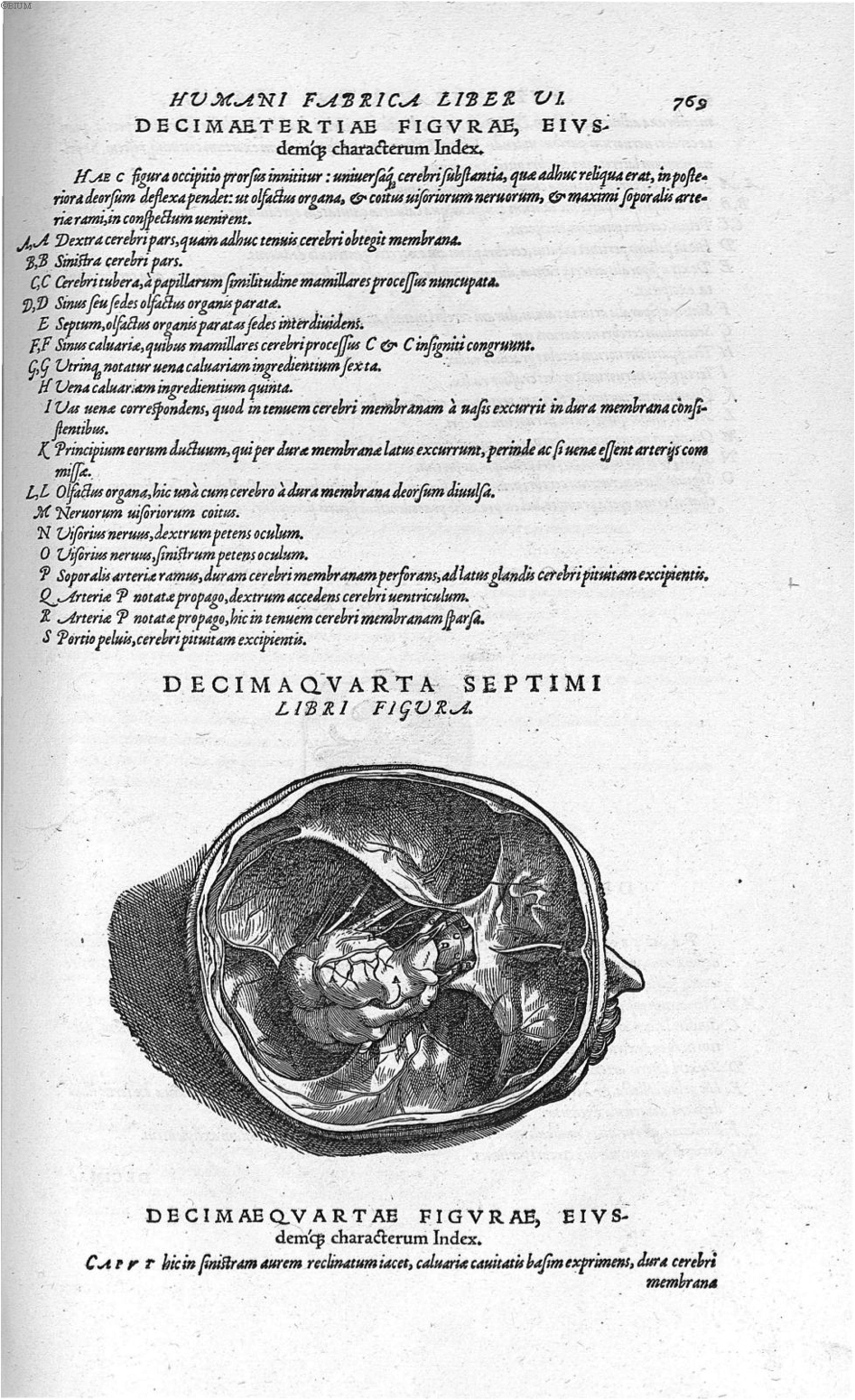
Figure 13. Vesalius, A., de Humani corporis fabrica Libri septem (p. 769), 1543. Link |
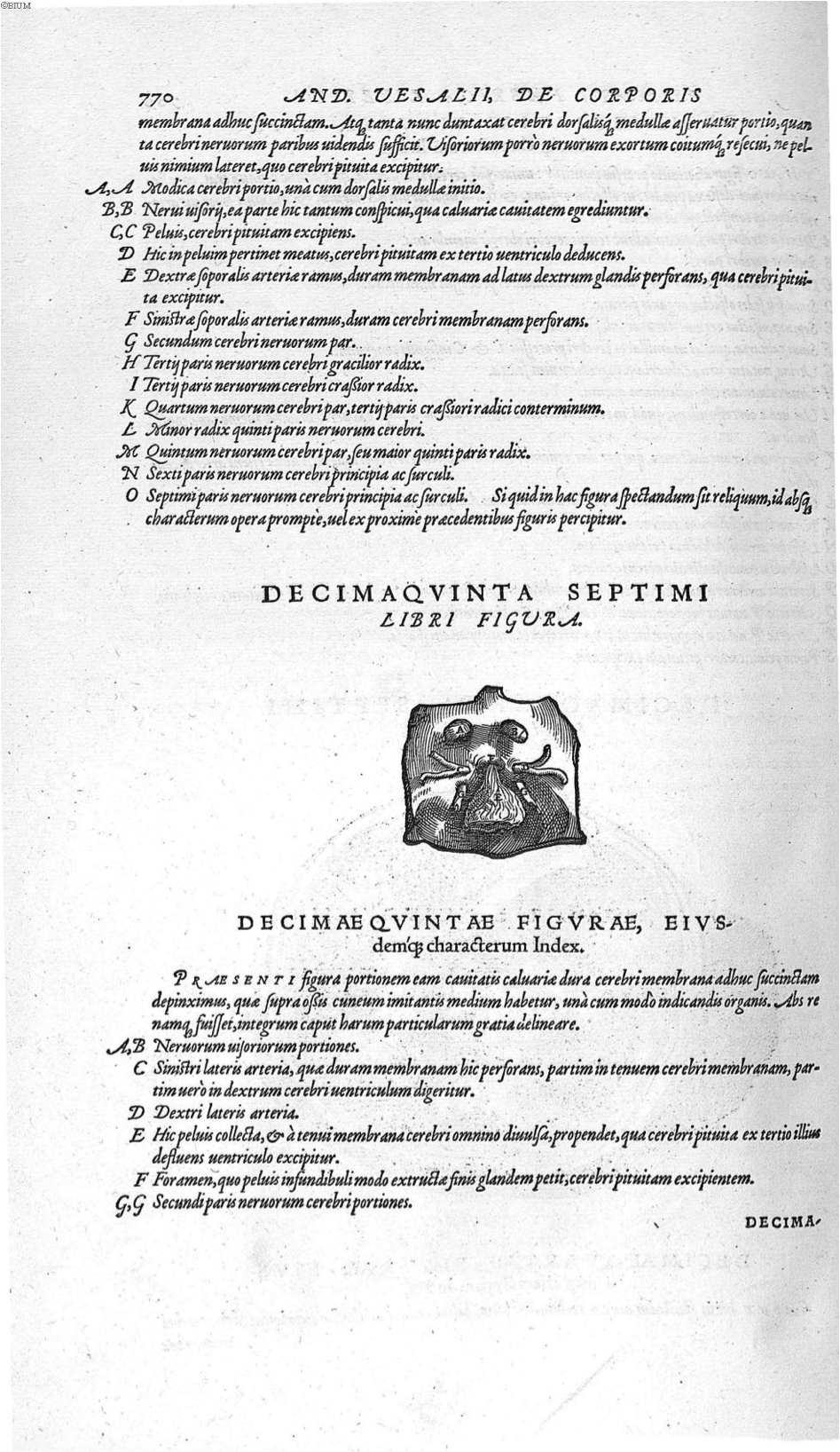
Figure 14. Vesalius, A., de Humani corporis fabrica Libri septem (p. 770), 1543. Link |
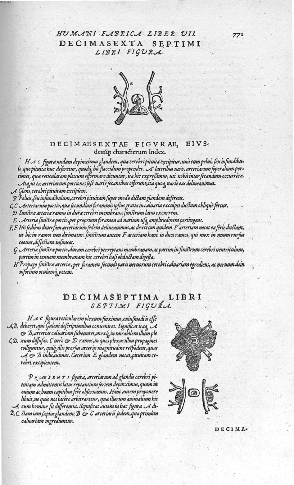
Figure 15. Vesalius, A., de Humani corporis fabrica Libri septem (p. 771), 1543. Link |
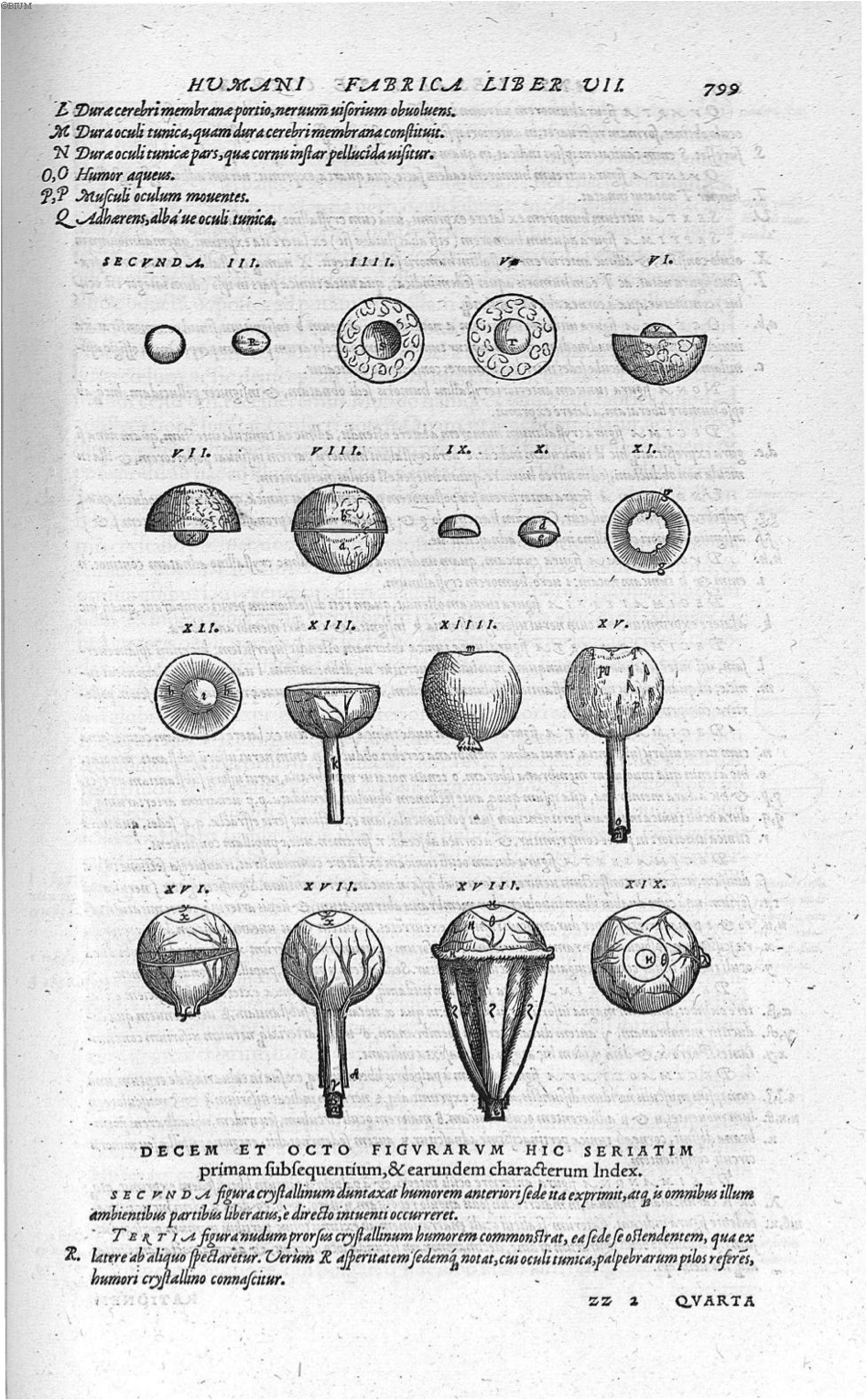
Figure 16. Vesalius, A., de Humani corporis fabrica Libri septem (p. 799), 1543. Link |
Willis T. (1664) Cerebri Anatome: Cui accessit nervorum descriptio et usus. Flesher, Martyn and Allestry; London.
Willis, T. (1667) Pathologiae cerebri et nervosi generis specimen. In quo Agitur de Morbis Convuslivis, et de Scorbuto. Excudebat Guil. Hall, impensierire Ja. Allestry; Oxford.
Willis, T. (1694) Opera omnia, nitidius quàm vnquam hactenus edita, plurimùm emendata, indice rerum copiosissimo, ac distinctione characterum exornata. Coloniae : Sumptibus G. Storti.

Figure 1. Willis, T., Opera omnia (p. x), 1694. Link |
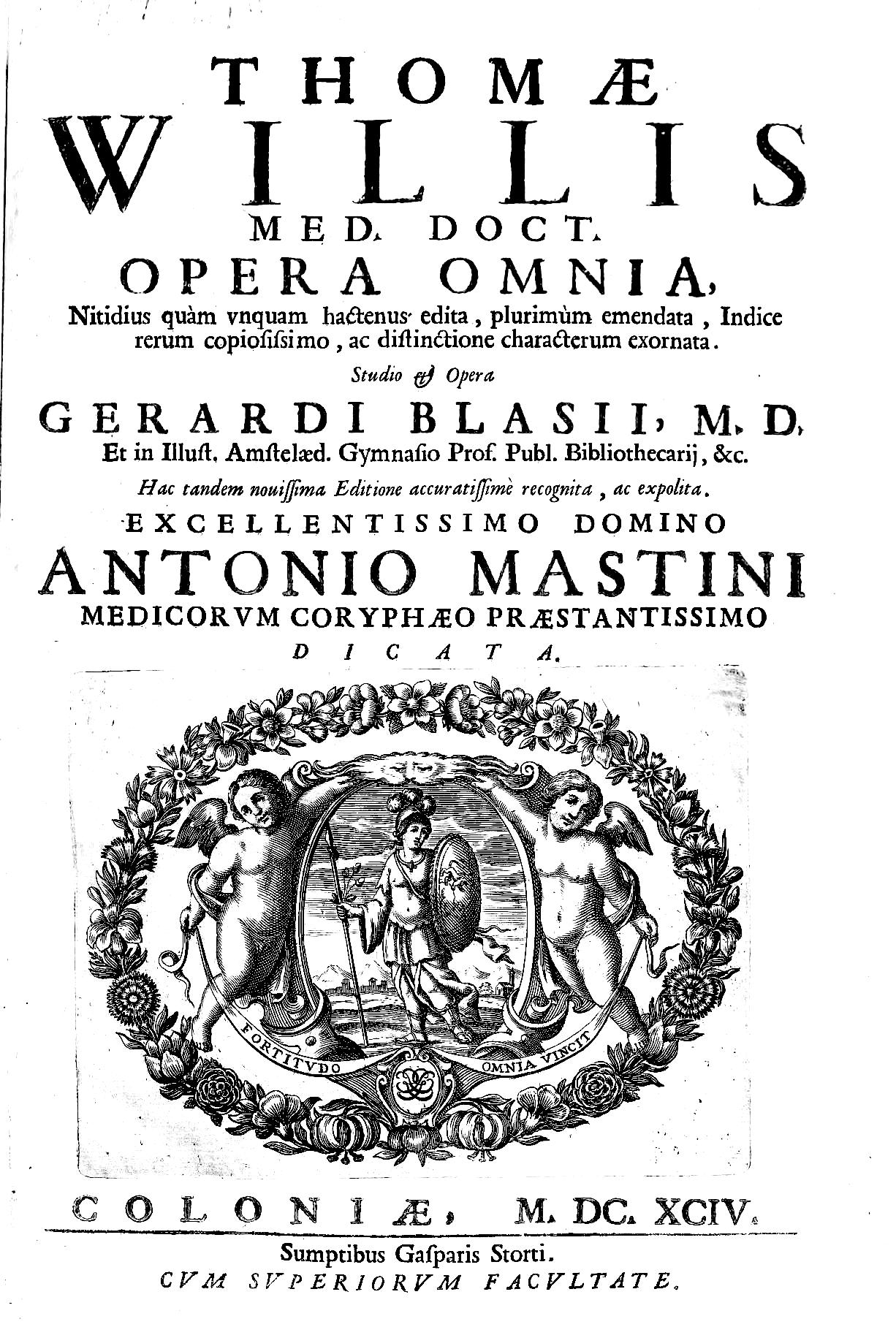
Figure 2. Willis, T., Opera omnia (page de couverture), 1694. Link |
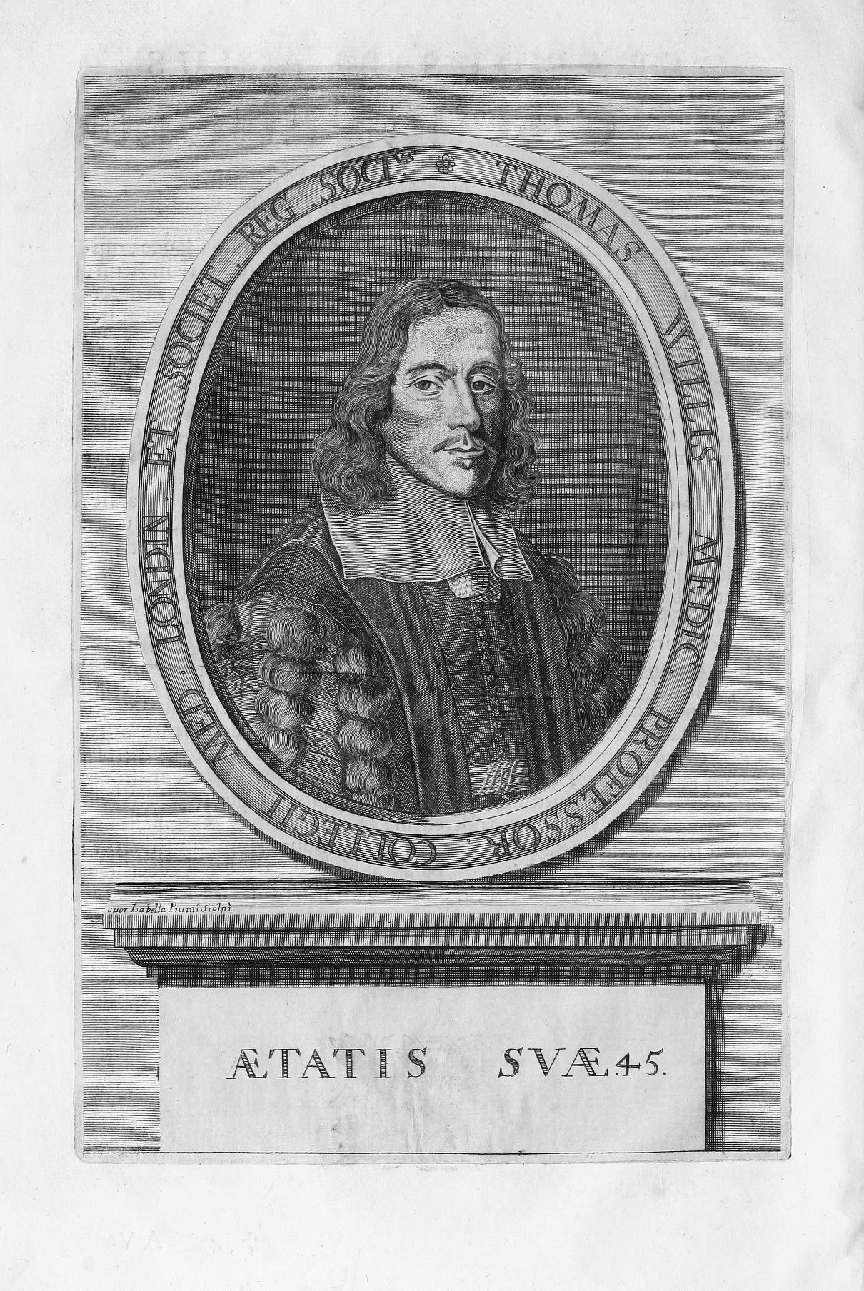
Figure 3. Willis, T., Opera omnia (p. i), 1694. Link |
Figure 4. Willis, T., Opera omnia (p. v), 1694. Link |
Figure 5. Willis, T., Opera omnia (p. vi), 1694. Link |
Figure 6. Willis, T., Opera omnia (p. vii), 1694. Link |
Figure 7. Willis, T., Opera omnia (p. viii), 1694. Link |
Figure 8. Willis, T., Opera omnia (p. ix), 1694. Link |
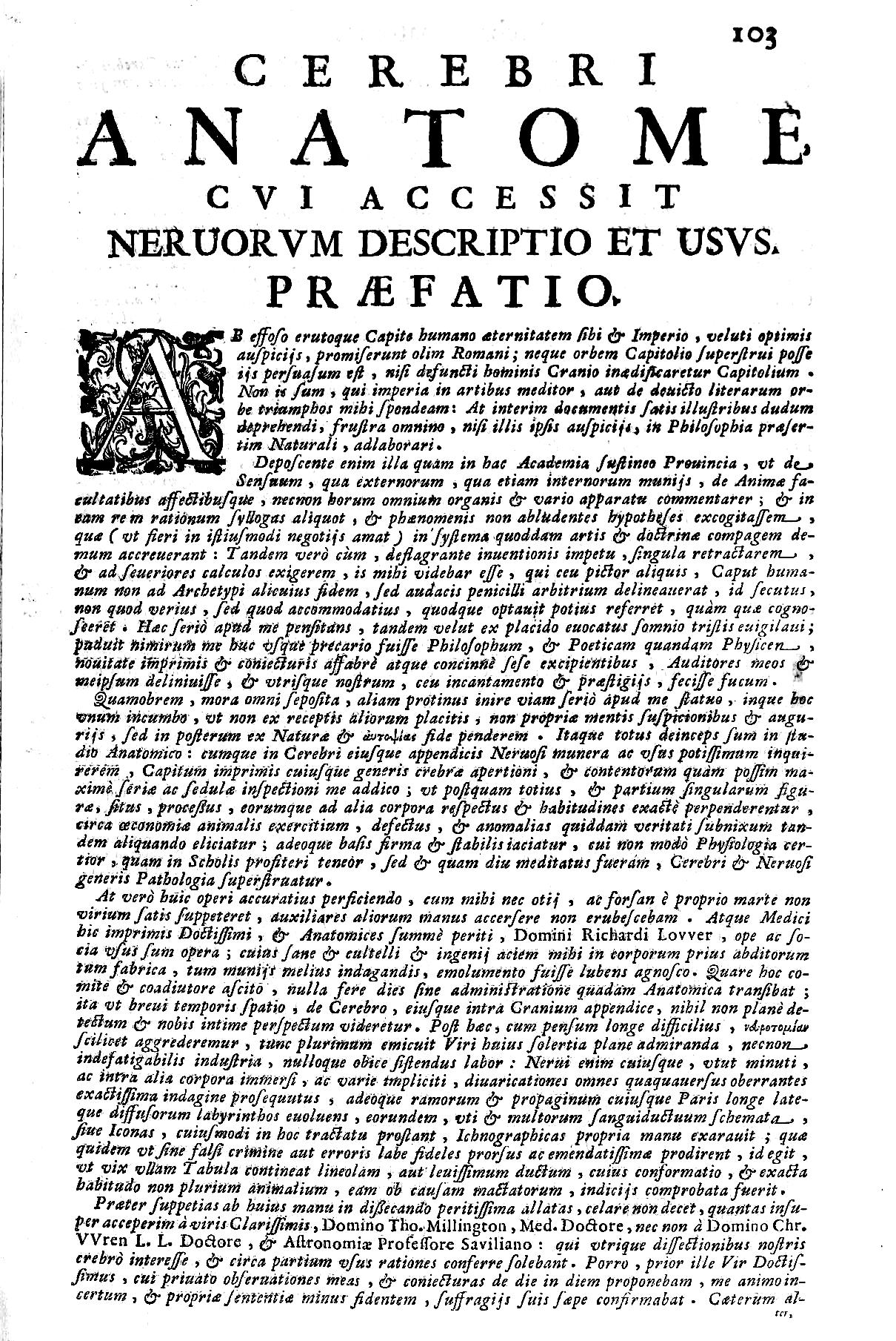
Figure 9. Willis, T., Opera omnia (p. 103), 1694. Link |
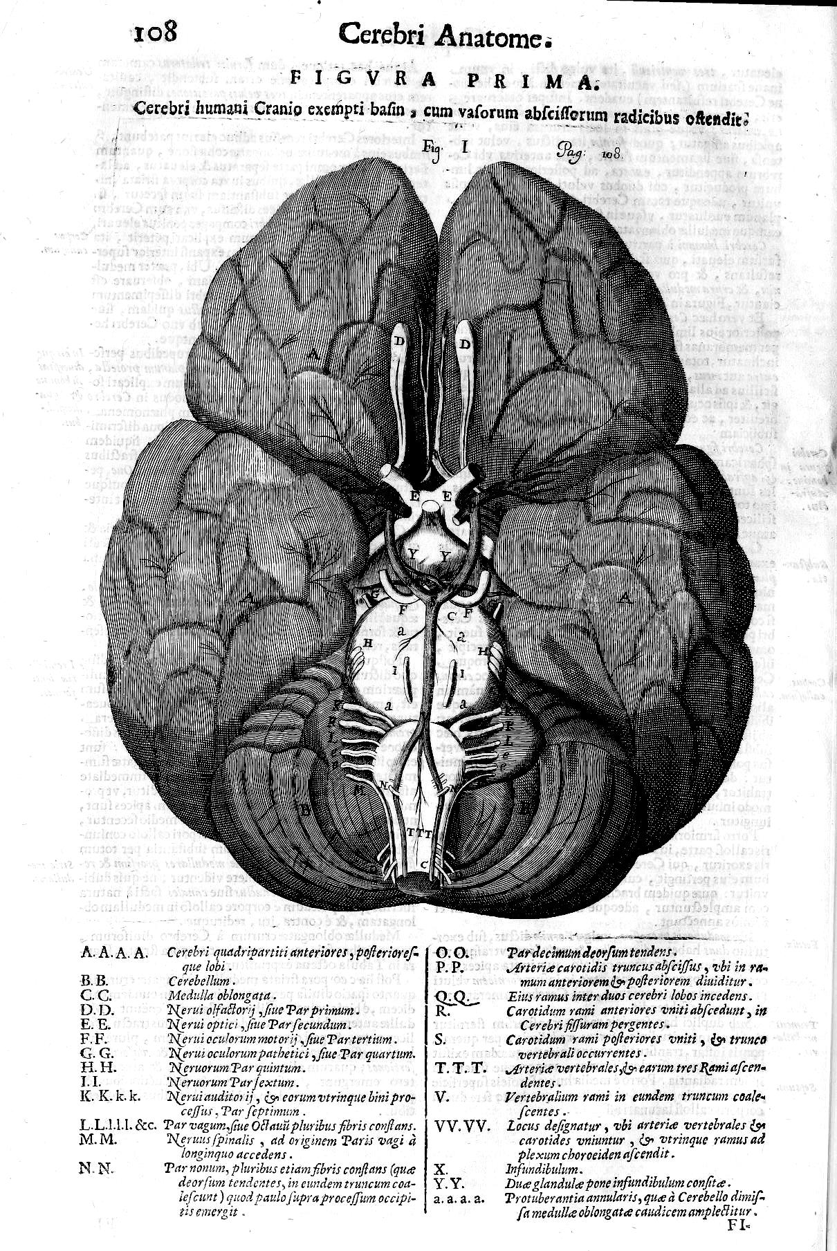
Figure 10. Willis, T., Opera omnia (p. 108), 1694. Link |
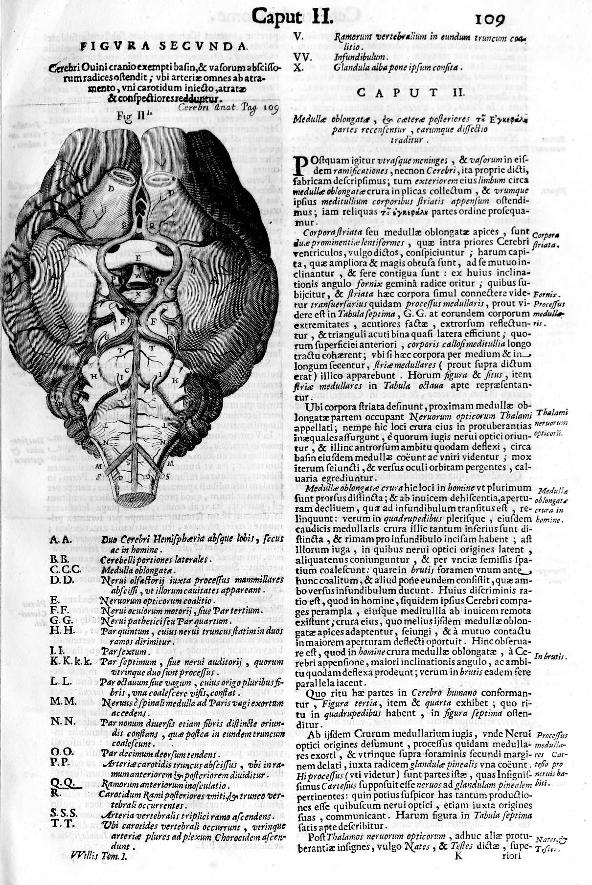
Figure 11. Willis, T., Opera omnia (p. 109), 1694. Link |
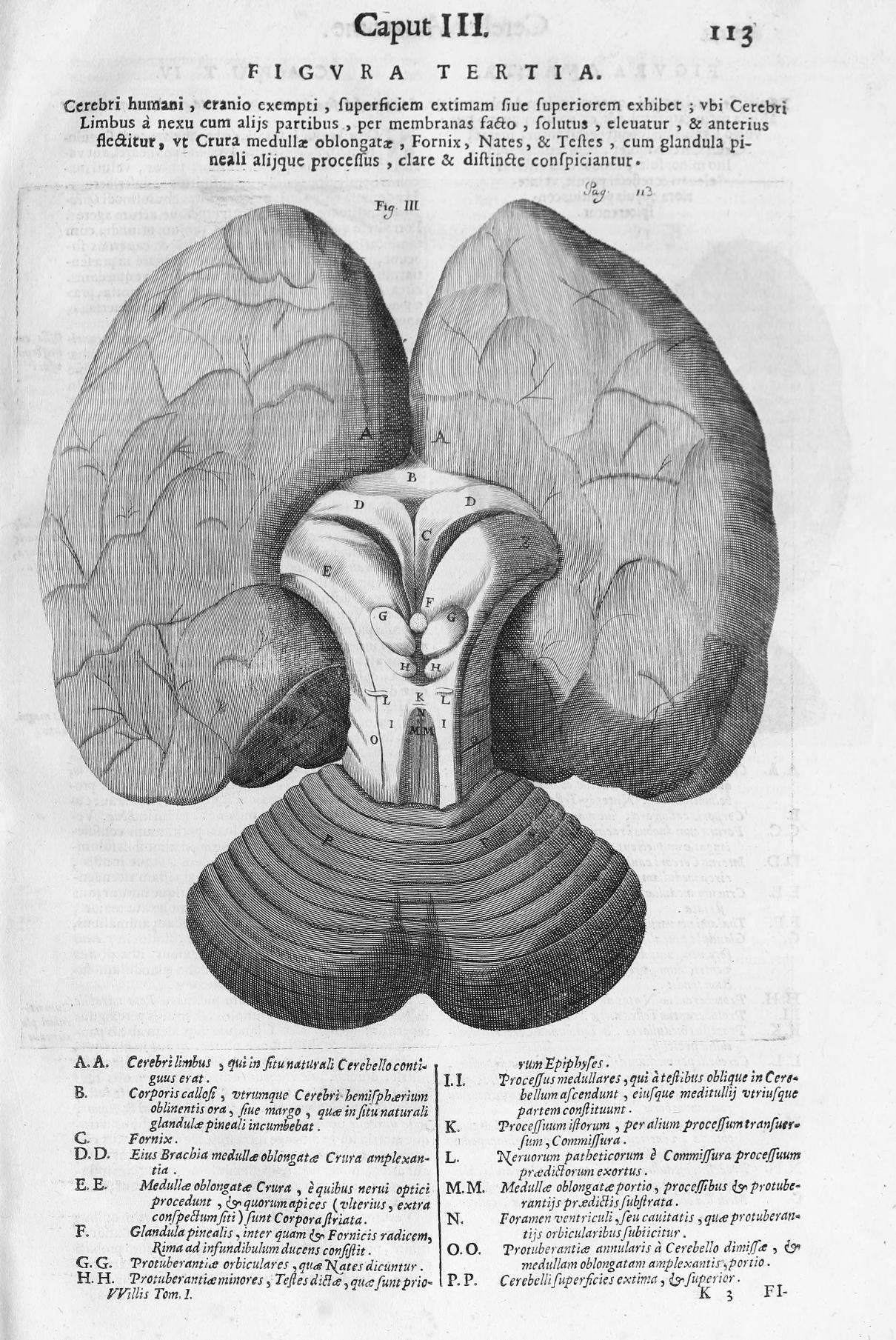
Figure 12. Willis, T., Opera omnia (p. 113), 1694. Link |
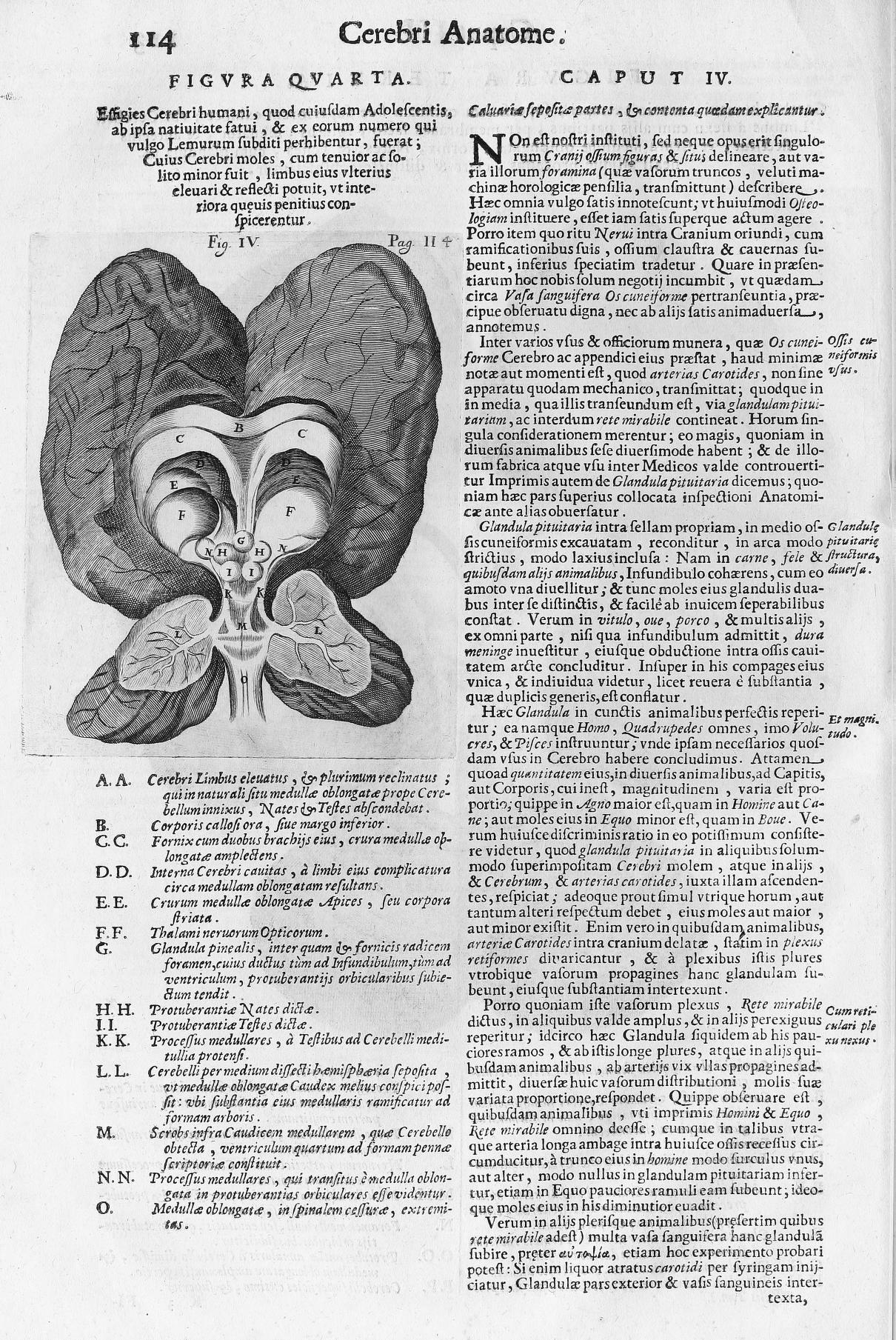
Figure 13. Willis, T., Opera omnia (p. 114), 1694. Link |
Figure 14. Willis, T., Opera omnia (p. 115), 1694. Link |
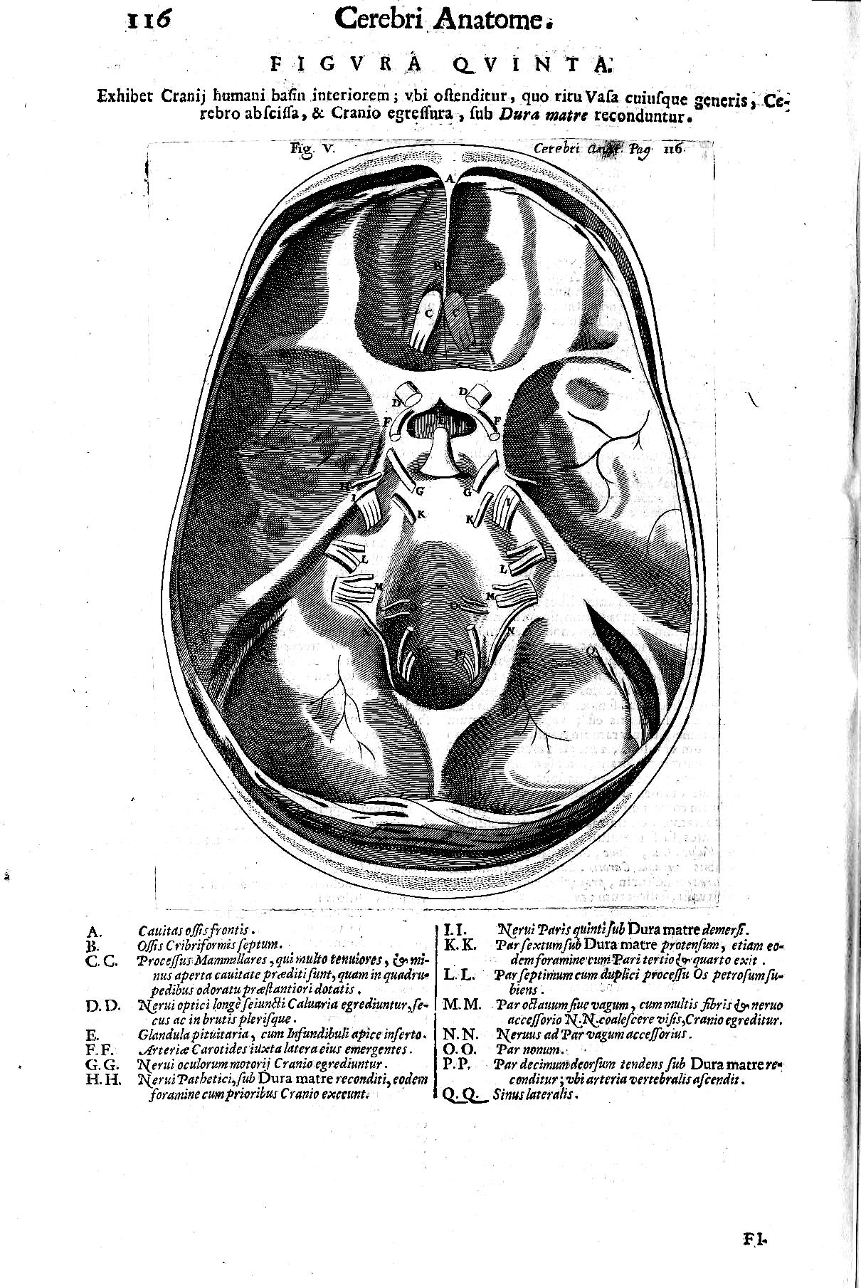
Figure 15. Willis, T., Opera omnia (p. 116), 1694. Link |
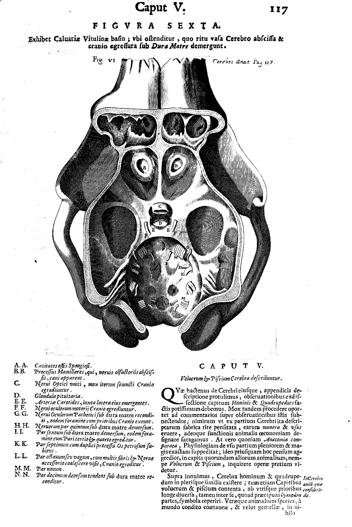
Figure 16. Willis, T., Opera omnia (p. 117), 1694. Link |
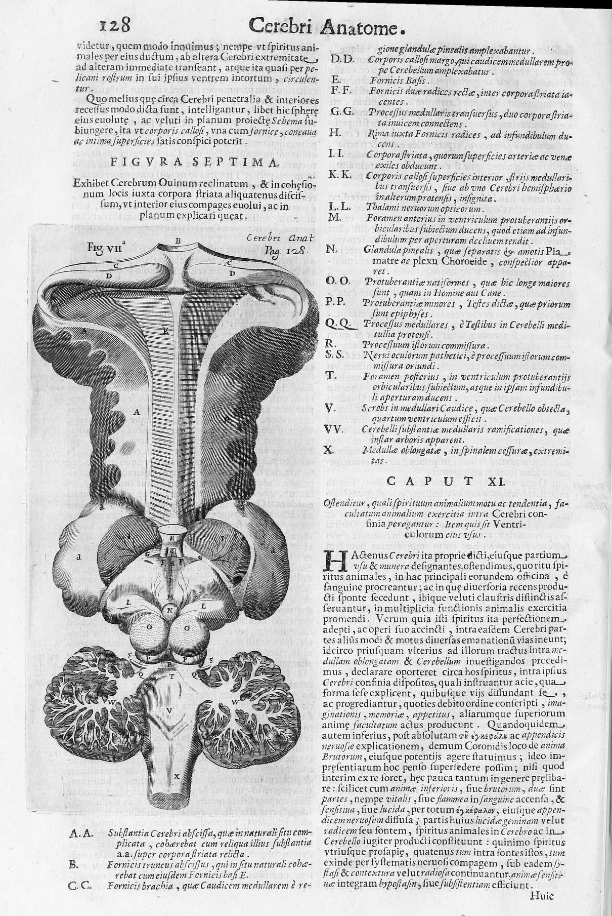
Figure 17. Willis, T., Opera omnia (p. 128), 1694. Link |
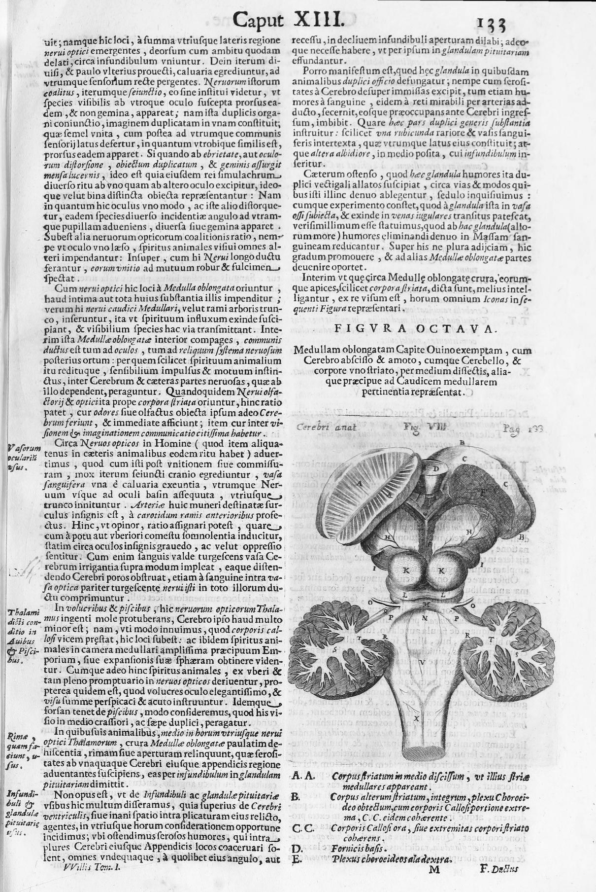
Figure 18. Willis, T., Opera omnia (p. 133), 1694. Link |
Figure 19. Willis, T., Opera omnia (p. 149), 1694. Link |
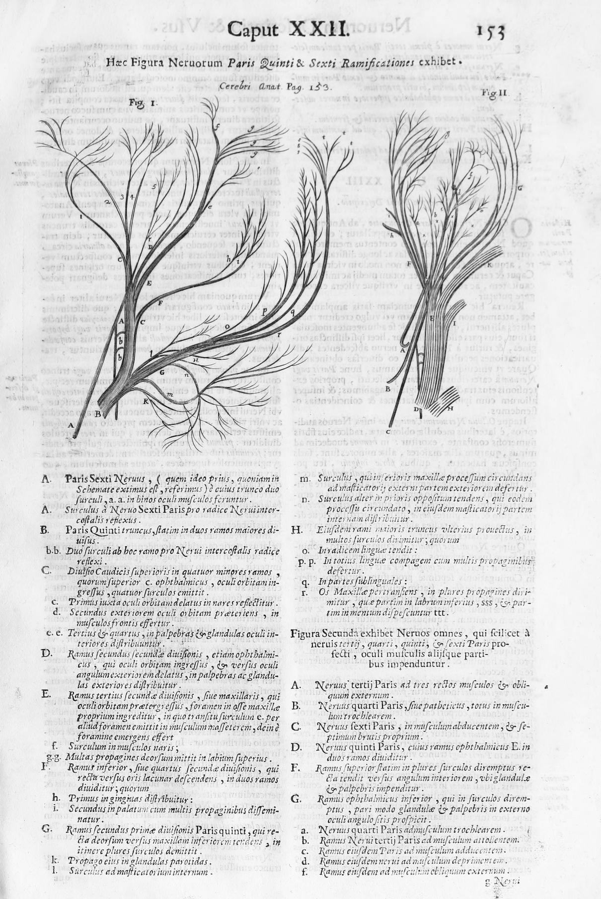
Figure 20. Willis, T., Opera omnia (p. 153), 1694. Link |
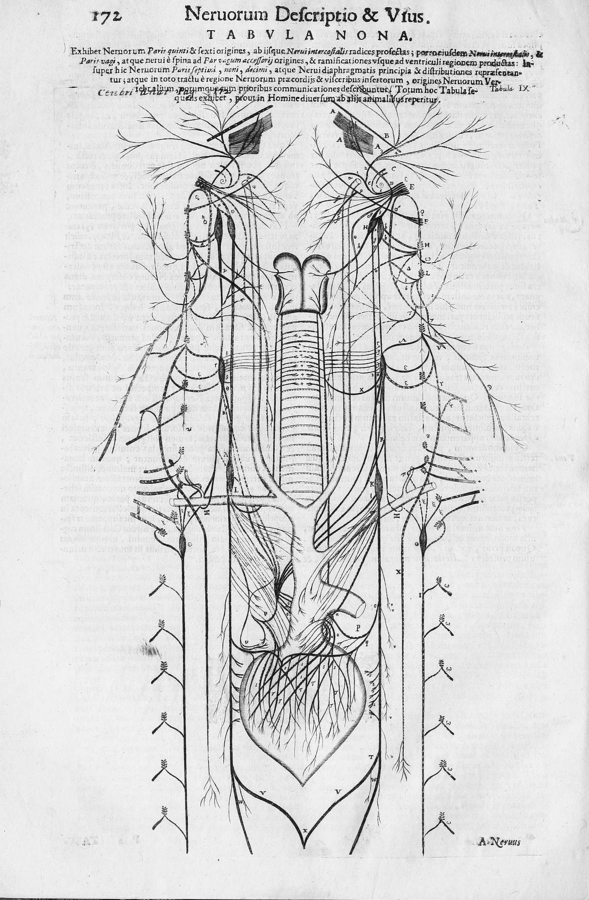
Figure 21. Willis, T., Opera omnia (p. 172), 1694. Link |
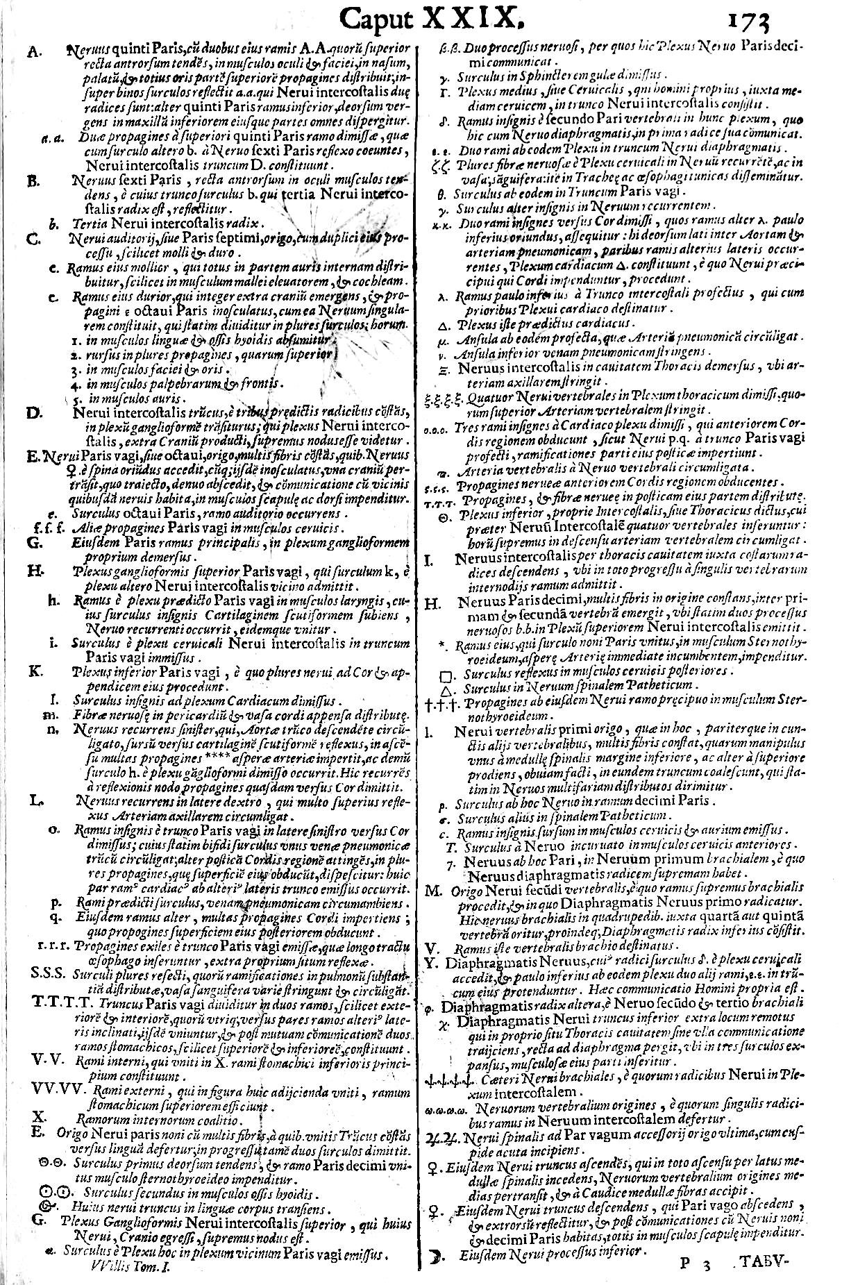
Figure 22. Willis, T., Opera omnia (p. 173), 1694. Link |
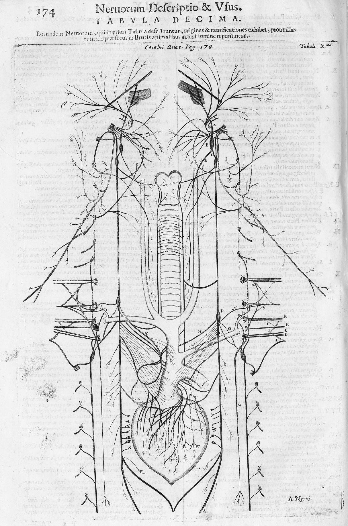
Figure 23. Willis, T., Opera omnia (p. 174), 1694. Link |
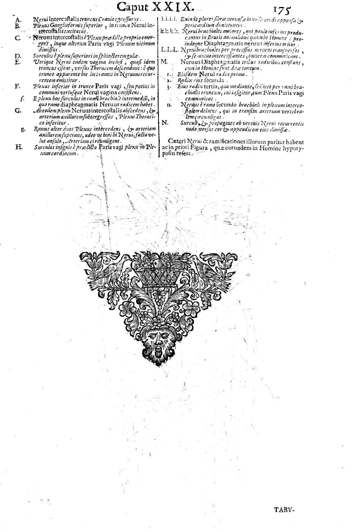
Figure 24. Willis, T., Opera omnia (p. 175), 1694. Link |
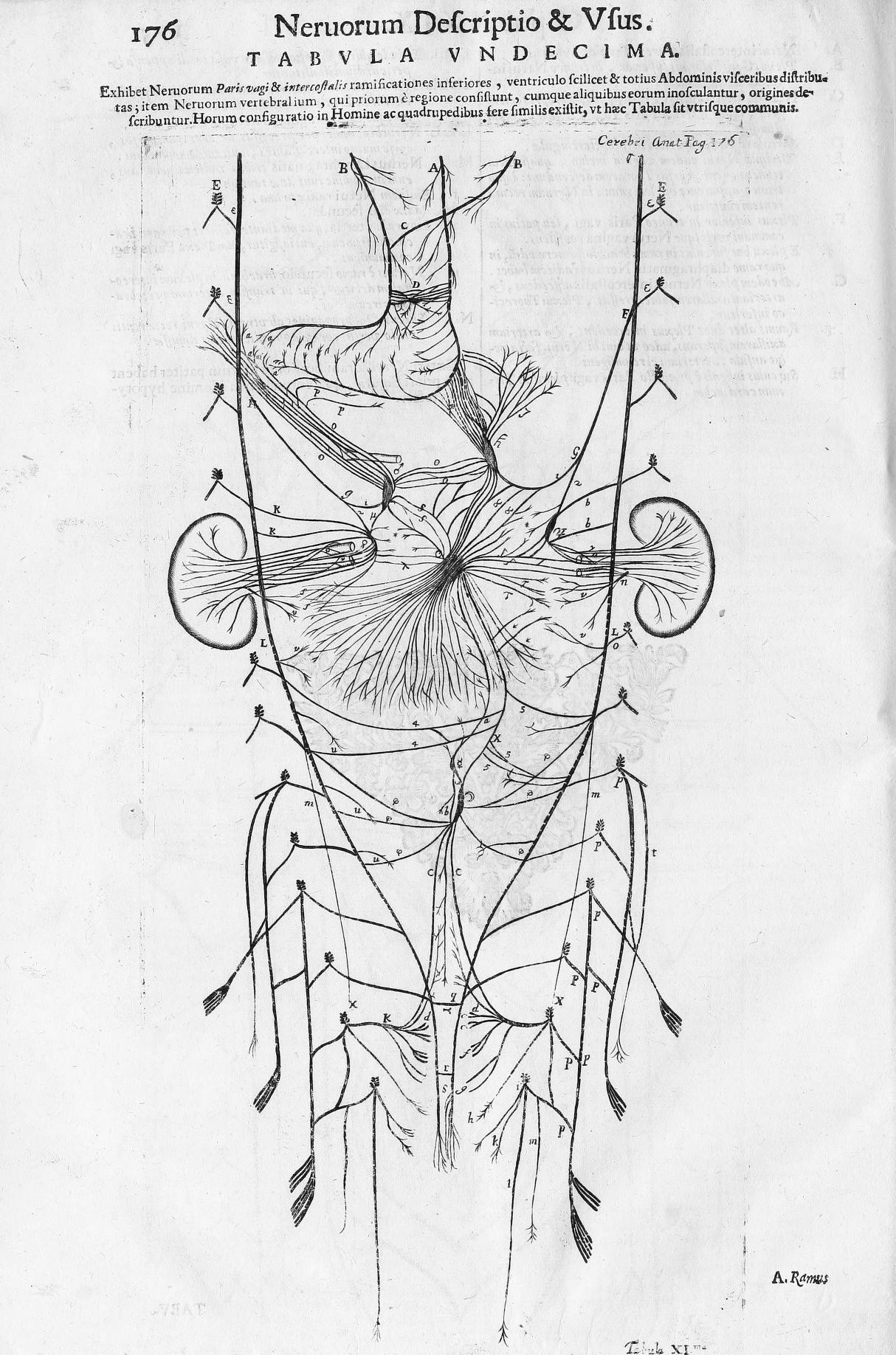
Figure 25. Willis, T., Opera omnia (p. 176), 1694. Link |
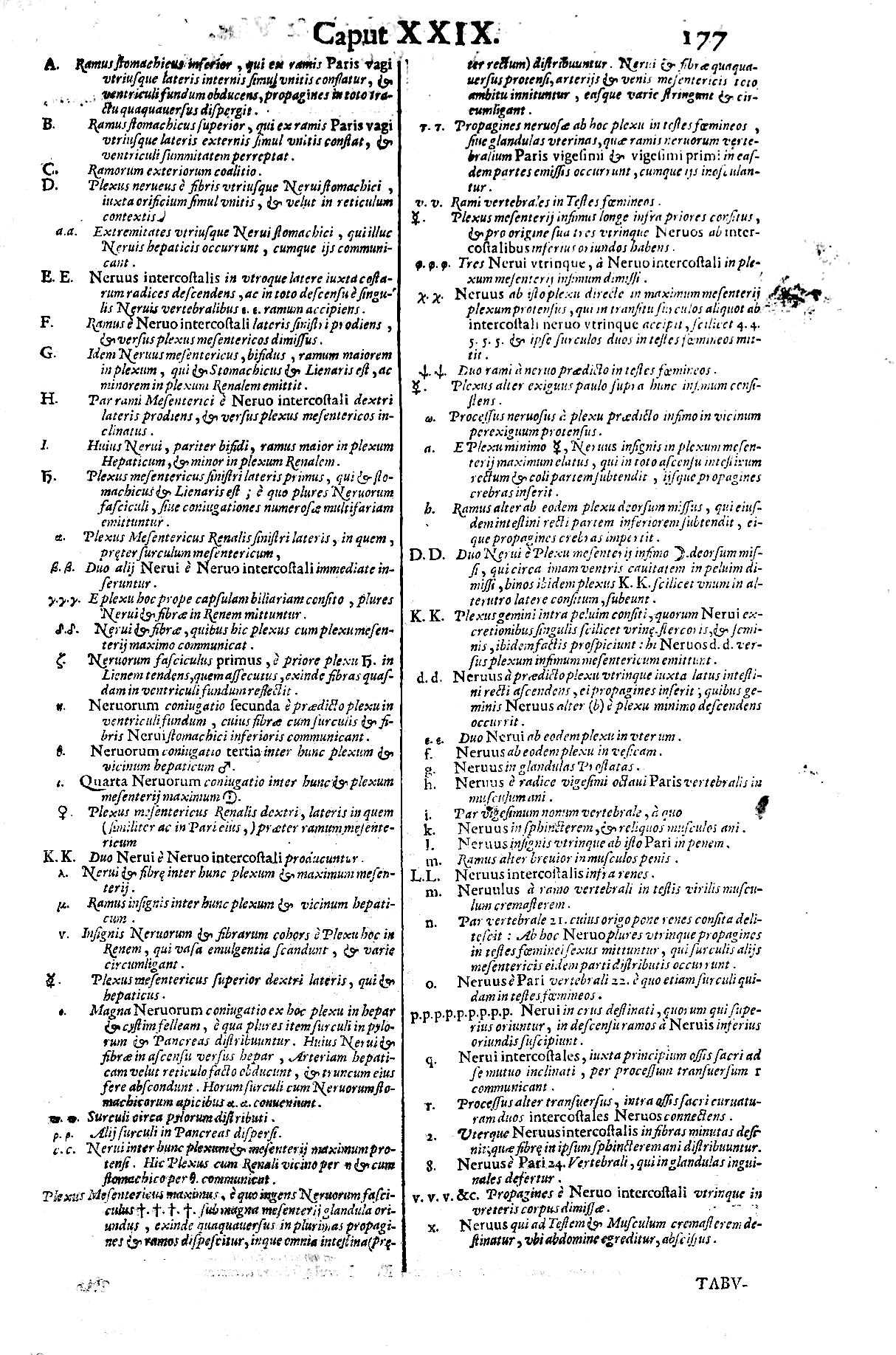
Figure 26. Willis, T., Opera omnia (p. 177), 1694. Link |
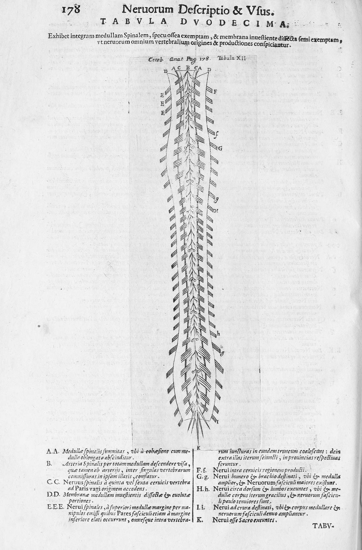
Figure 27. Willis, T., Opera omnia (p. 178), 1694. Link |
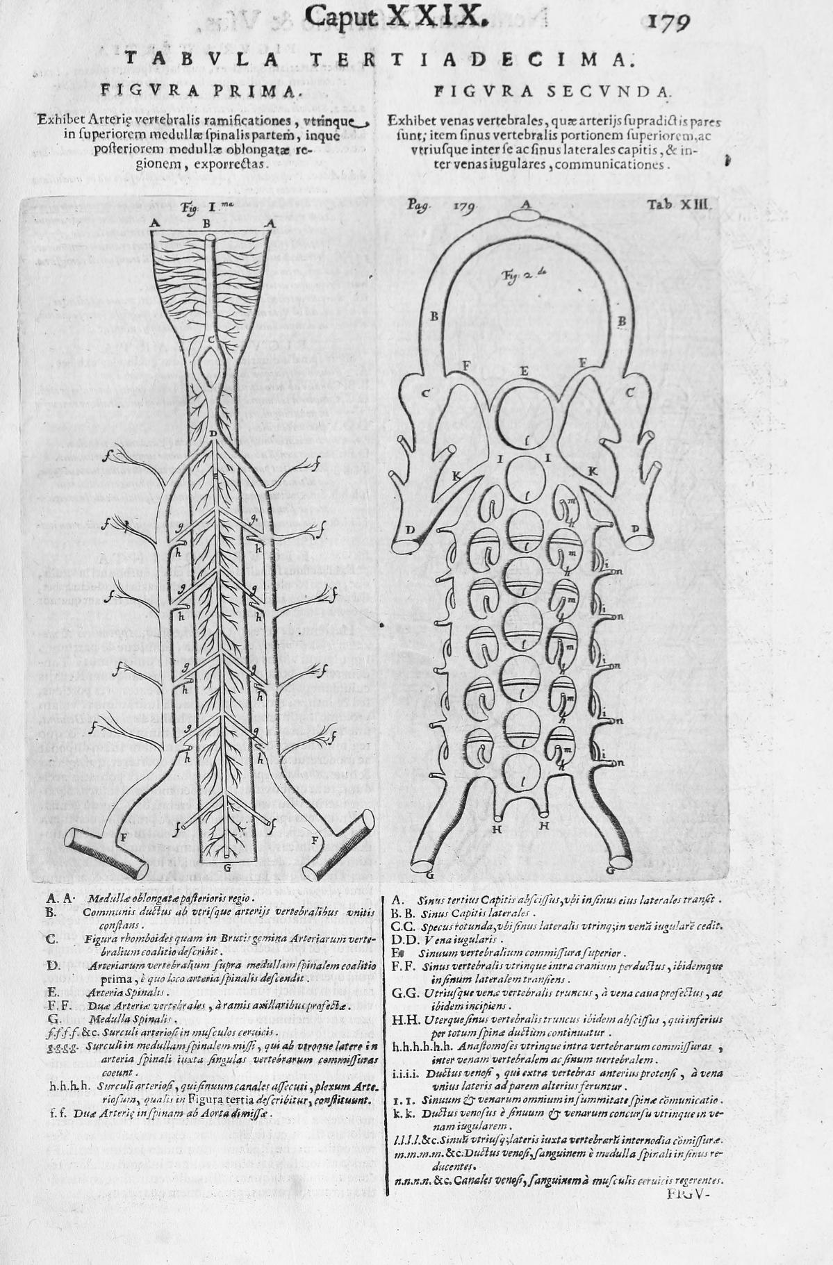
Figure 28. Willis, T., Opera omnia (p. 179), 1694. Link |
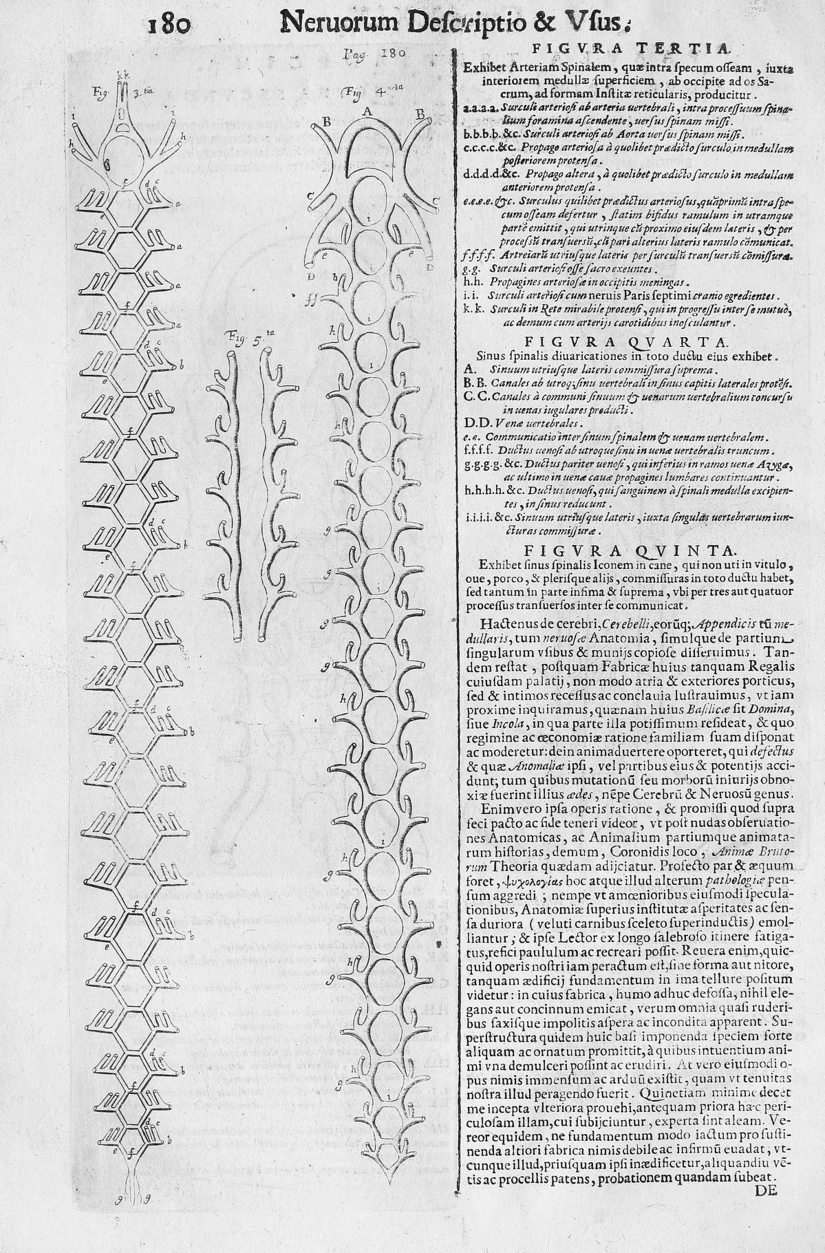
Figure 29. Willis, T., Opera omnia (p. 180), 1694. Link |
Figure 30. Willis, T., Opera omnia (p. 181), 1694. Link |
Figure 31. Willis, T., Opera omnia (p. 189), 1694. Link |
Figure 32. Willis, T., Opera omnia (p. 294), 1694. Link |
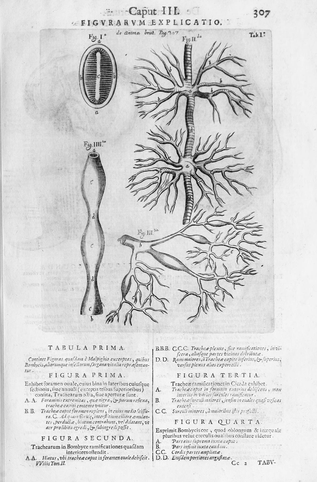
Figure 33. Willis, T., Opera omnia (p. 307), 1694. Link |
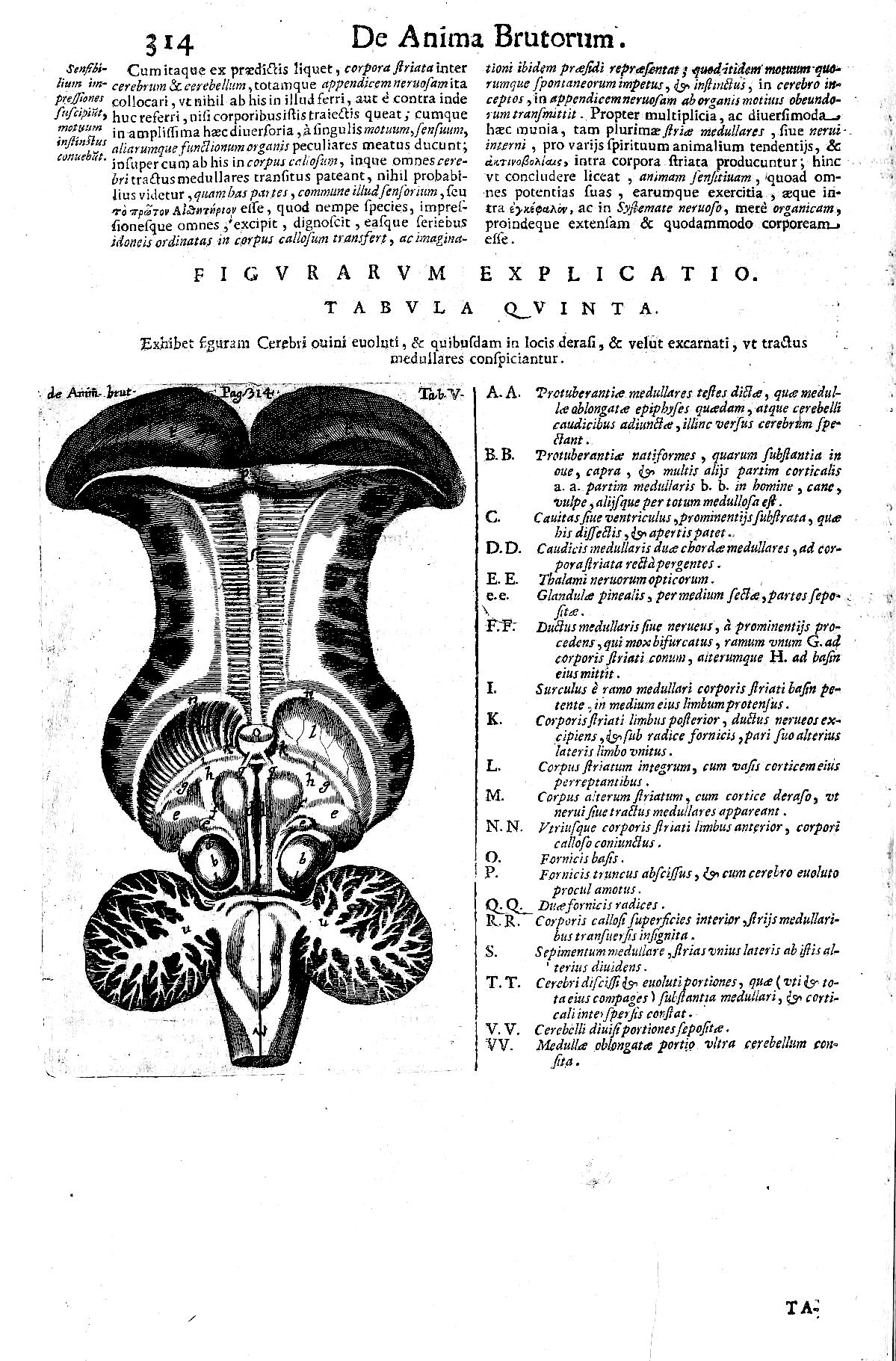
Figure 34. Willis, T., Opera omnia (p. 314), 1694. Link |
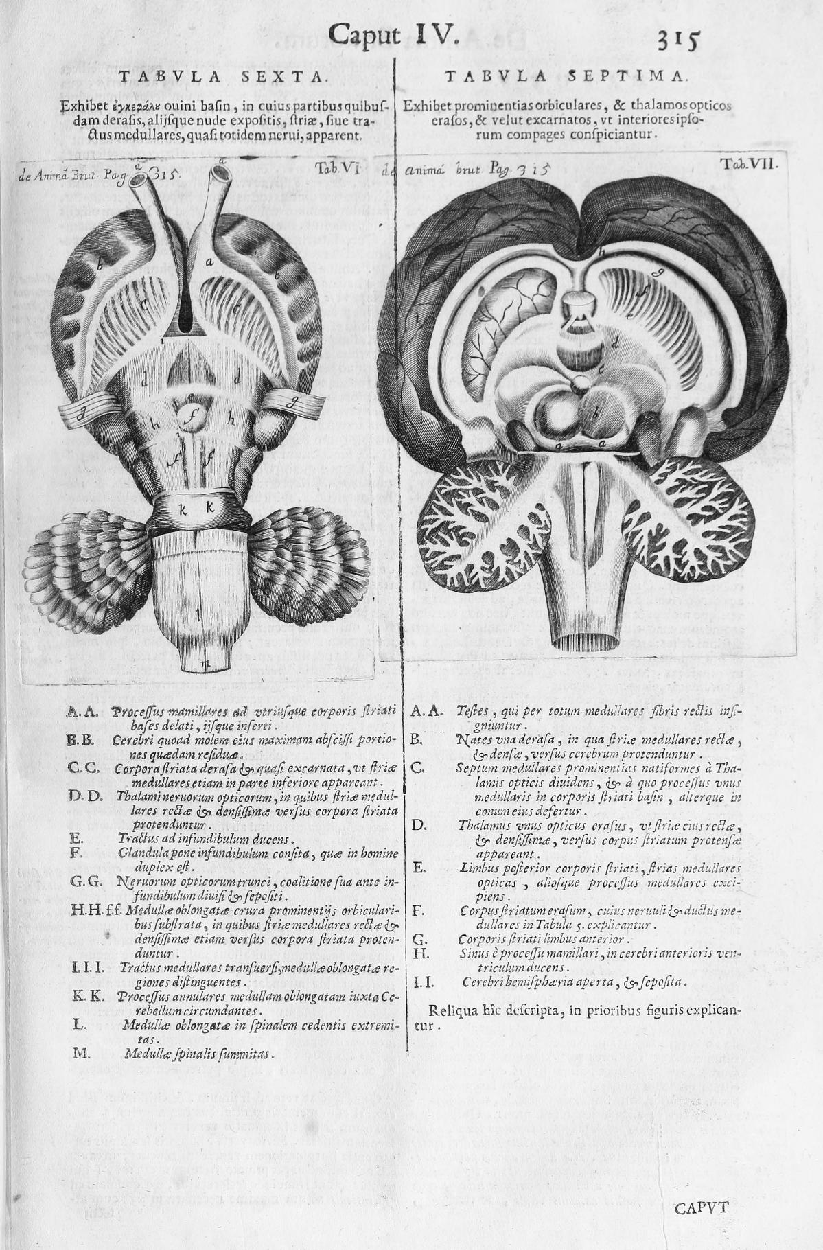
Figure 35. Willis, T., Opera omnia (p. 315), 1694. Link |
Figure 36. Willis, T., Opera omnia (p. 324), 1694. Link |
Galvani L (1791) De viribus electricitatis in motu musculari commentarius. De Bononiensi Scientiarum et Artium Instituto atque Academia commentarii 7: 363–418.
|
Figure 1. Neurocranium (computer animation N°0). Link |
Figure 2. Facial bones (computer animation n°1). Link |
Figure 3. Facial bones and Cranial bones (computer animation N°1). Link |
Figure 4. Facial bones and Cranial bones (computer animation N°2). Link |
|
Figure 1. Neurocranium (computer animation N°0). Link |
Figure 2. Facial bones (computer animation n°1). Link |
Figure 3. Facial bones and Cranial bones (computer animation N°1). Link |
Figure 4. Facial bones and Cranial bones (computer animation N°2). Link |
Golgi, C. (1873) Sulla struttura della sostanza grigia del cervelo. Gazzetta Medica Italiana. Lombardia 33, 244–246. Golgi C. (1873) On the structure of the grey matter of the brain. (Translated by M. Santini). In Golgi Centenmial Symposium (1975, M. Santini, Ed.). New York : Raven, pp. 647-650.
|
Figure 1. Neurocranium (computer animation N°0). Link |
Figure 2. Facial bones (computer animation n°1). Link |
Figure 3. Facial bones and Cranial bones (computer animation N°1). Link |
Figure 4. Facial bones and Cranial bones (computer animation N°2). Link |
|
Figure 1. Neurocranium (computer animation N°0). Link |
Figure 2. Facial bones (computer animation n°1). Link |
Figure 3. Facial bones and Cranial bones (computer animation N°1). Link |
Figure 4. Facial bones and Cranial bones (computer animation N°2). Link |
Waldeyer-Hartz, Wilhelm von (1891). Das Gibbongehirn. Internationale Beiträge zur wissenschaftlichen Medizin 1: 1-40.
|
Figure 1. Neurocranium (computer animation N°0). Link |
Figure 2. Facial bones (computer animation n°1). Link |
Figure 3. Facial bones and Cranial bones (computer animation N°1). Link |
Figure 4. Facial bones and Cranial bones (computer animation N°2). Link |
|
Figure 1. Neurocranium (computer animation N°0). Link |
Figure 2. Facial bones (computer animation n°1). Link |
Figure 3. Facial bones and Cranial bones (computer animation N°1). Link |
Figure 4. Facial bones and Cranial bones (computer animation N°2). Link |